Non-Destructive Quantification of Cytotoxicity in Live HeLa Cells
10 Dec 2018Cytotoxicity assays are a crucial step in screening for and developing therapeutic anti-cancer drugs. Most assays designed to measure cytotoxicity in vitro evaluate cell membrane integrity or metabolic activity after exposure, but are typically based on studying a single time point and require disturbing the growth of cells in culture. This application note demonstrates an automated, non-destructive method to monitor and quantify cytotoxicity based on its effects on confluency.
Related products
Request Quote for All Products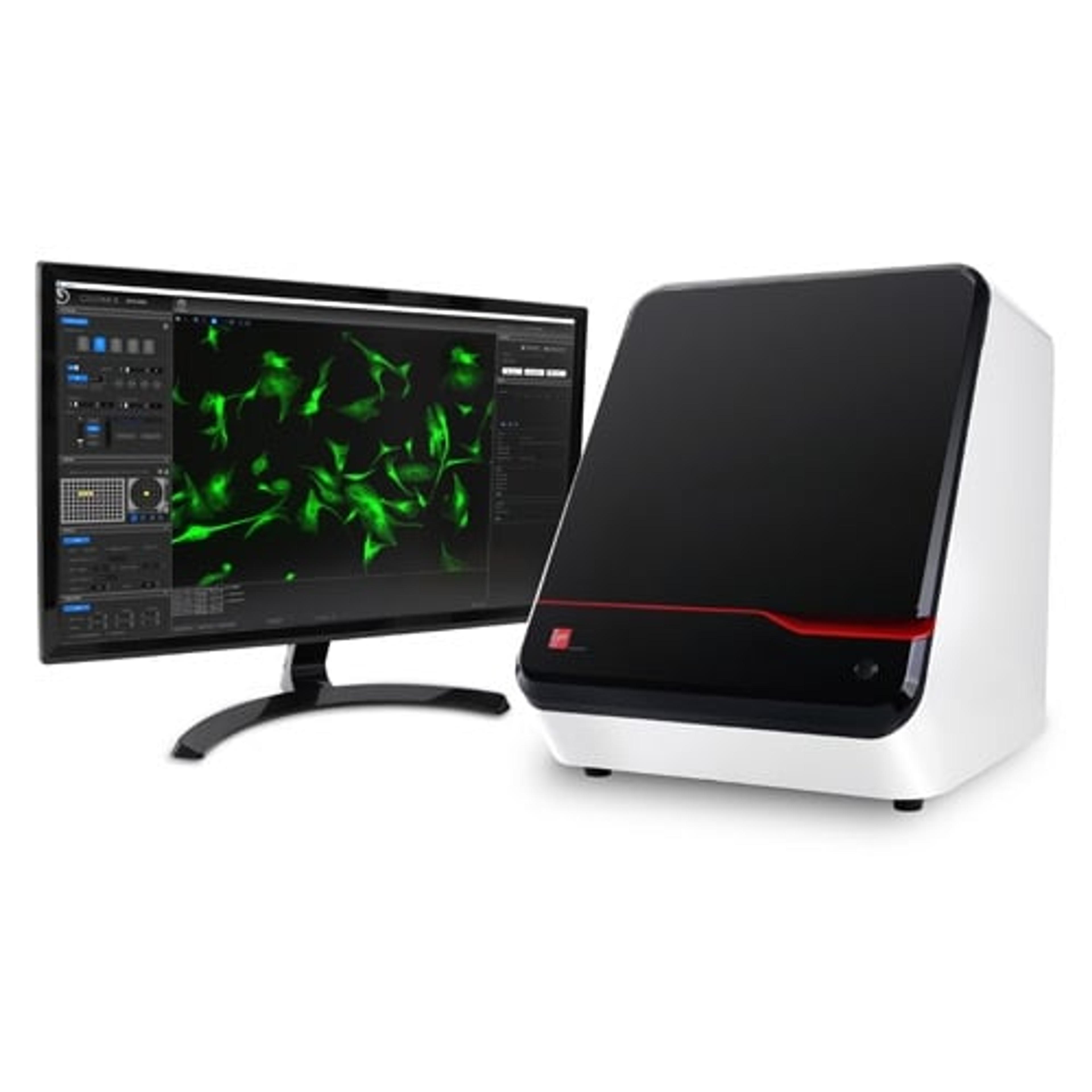
CELENA® X High Content Imaging System
Logos BiosystemsThe CELENA ® X High Content Imaging System is an integrated imaging system designed for rapid, high content image acquisition and analysis.
Links
Tags
High-Content ScreeningHigh-content screening (HCS), also known as high-content analysis (HCA), is a high-throughput technique used in drug discovery to identify substances that alter the phenotype of cells. HCS uses fluorescent microscopic imaging and automated image analysis to investigate cellular events such as apoptosis, cell viability, GPCR activation, oxide production, neurite outgrowth, and cell signaling. Find the best fluorescent labeling reagents, cellular assays, and high-content imaging systems in our peer-reviewed product directory: compare products, check customer reviews and receive pricing direct from manufacturers.In VitroIn vitro refers to experiments conducted outside living organisms, often in controlled lab environments such as petri dishes or test tubes. In vitro models are widely used in drug testing, cell biology, and disease research. Explore in vitro research tools in our peer-reviewed product directory; compare products, check reviews, and get pricing directly from manufacturers.CytotoxicityCytotoxicity assays measure the toxic effects of substances on cells, often used in drug testing and environmental studies. These tests are crucial in determining the safety of chemicals and pharmaceutical compounds. Explore cytotoxicity testing tools in our peer-reviewed product directory; compare products, check reviews, and get pricing directly from manufacturers.High Content ImagingHigh content imaging is a method combining two or more fluorescent microscopy experiments to identify substances that alter a cell’s phenotype in a desired manner. The process is adapted to multi-well plates and both the image acquisition and analysis are automated.Drug Discovery & Development Screening