Products & ReviewDrug discovery > Target Discovery
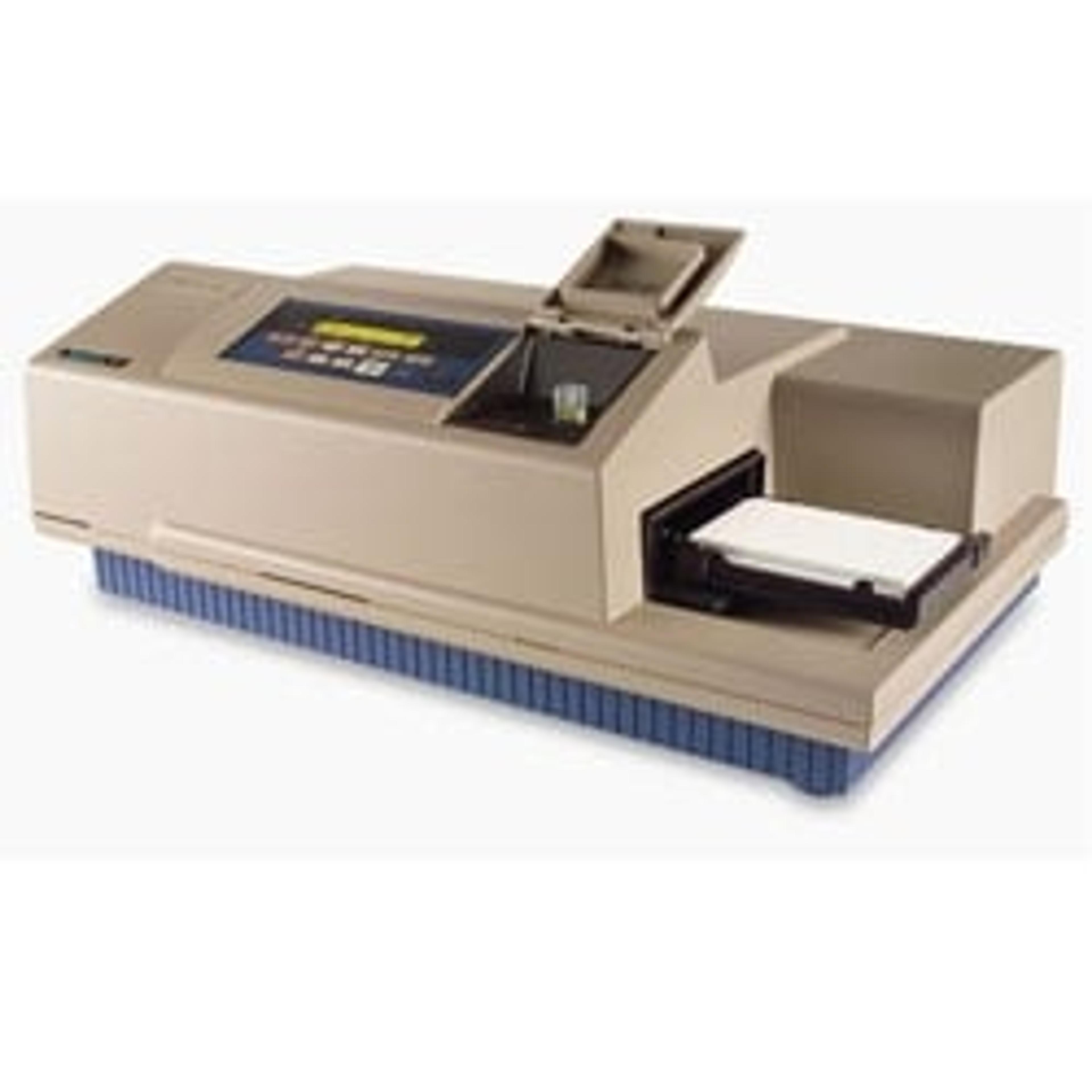
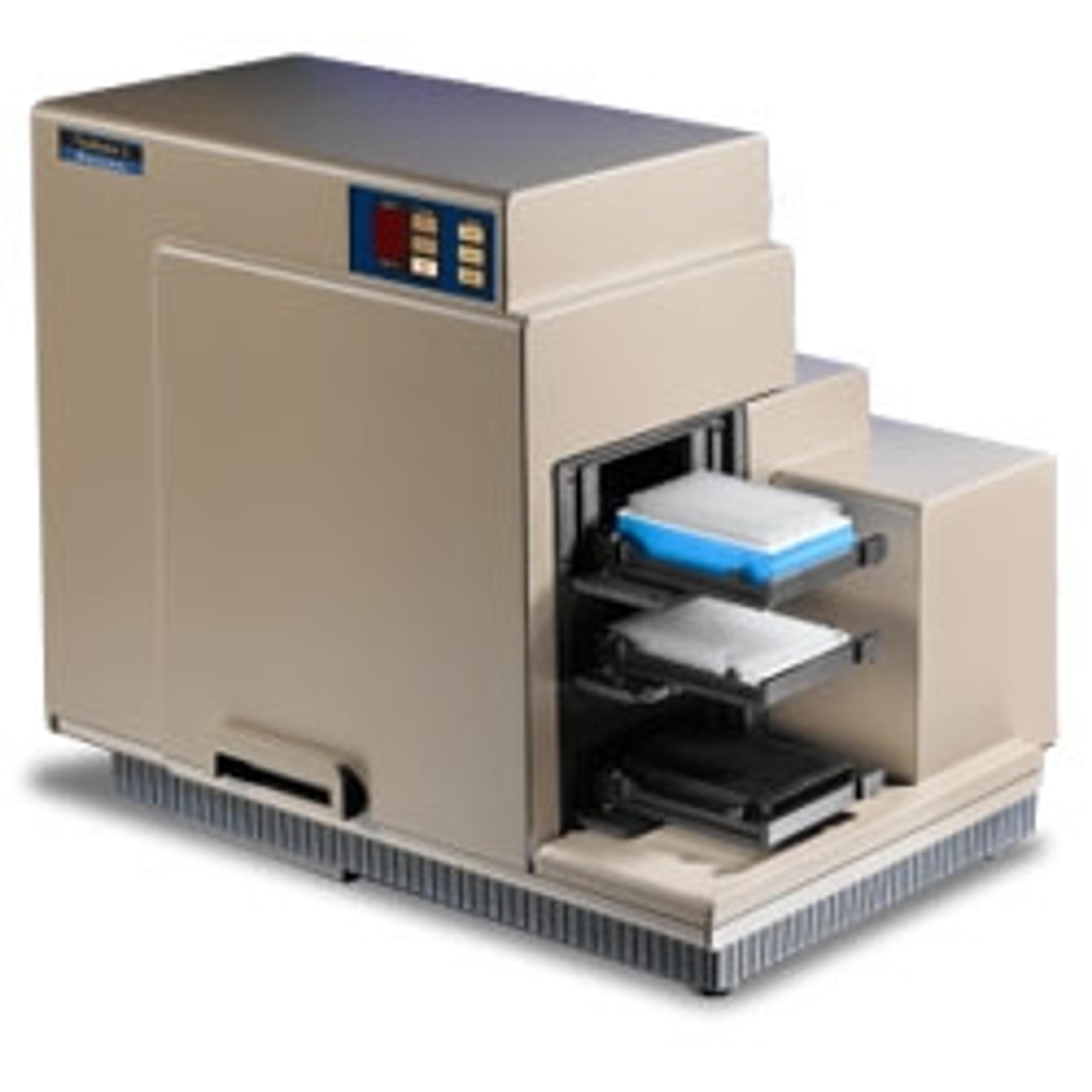
CloneSelect Imager
Label-free imaging solution for assurance of monoclonality


Request Pricing
Receive your quote directly from the manufacturer.
Description
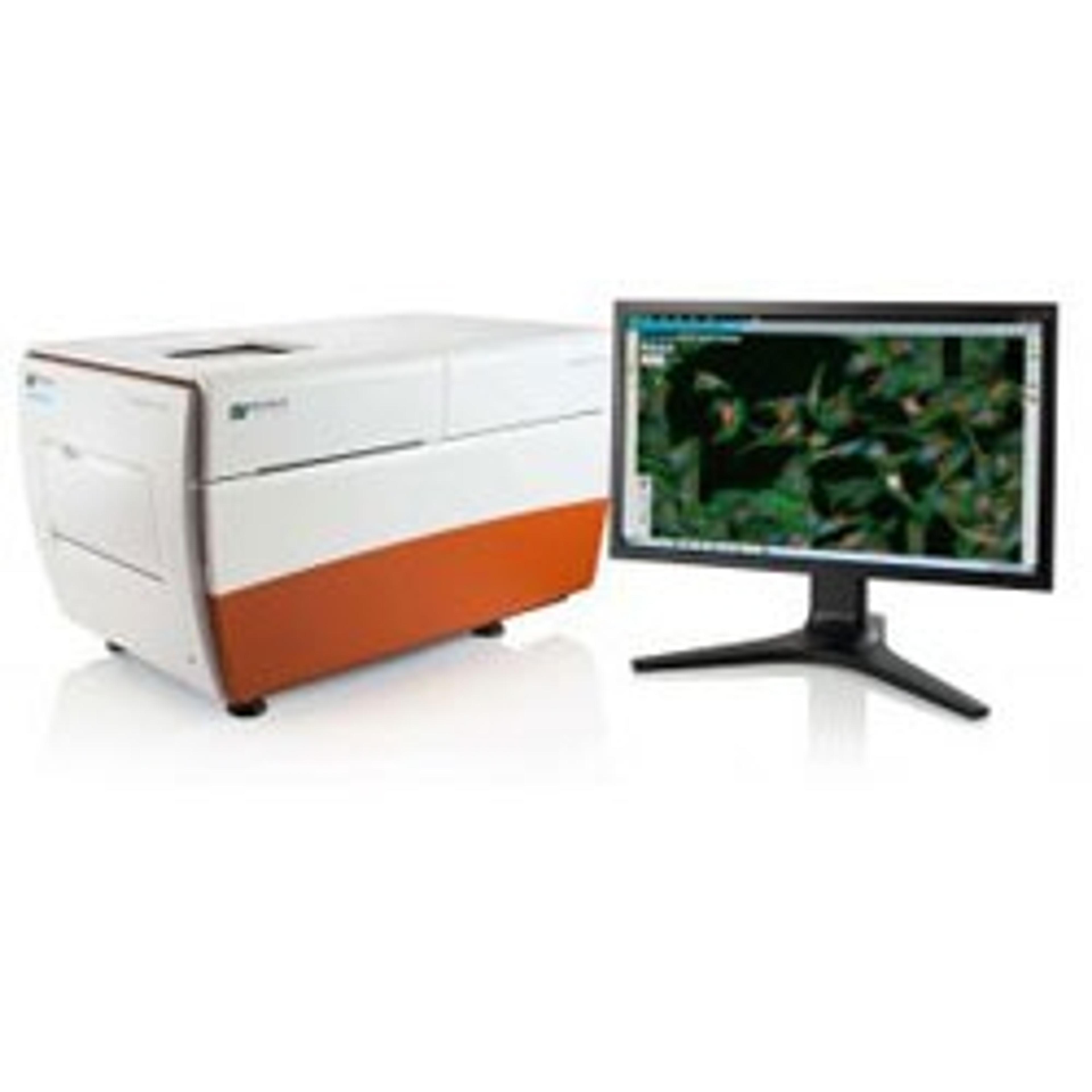
The CloneSelect™ Imager is a high-throughput automated system for imaging and analyzing mammalian colony formation and confluence. Tracking the growth of a colony from a single cell is effortless as barcoded plates are tracked over time. Automated fluorescence or white light image acquisition and analysis provide accurate, objective, and consistent results. With the ability to image a 96-well plate in 90 seconds, the imager will increase throughput and productivity.
KEY BENEFITS
- Assess cell confluence objectively and quantitatively
- Streamline workflow: image, analyze, report
- Image every well anytime to track colony formation and verify monoclonality
Track and record cell growth
- Automatically scans every well in every plate. Algorithms are optimized for accurate cell detection and address varying cell types and conditions.
- Generates growth curves for each well
- Enables viewing and tracking of every well in every plate
- Reveals additional information on cellular morphology and an understanding of growth characteristics
Verify monoclonality
- Seed one cell per well and image at any time point
- Focus on wells with a single loci of growth and view image history to verify monoclonality
- Verify colony origin by tracking image history of each well. The optional Monoclonality Report feature automatically organizes supporting image evidence into an easily shareable report
Colony formation assay
- After seeding in a matrix that enhances colony formation, image every well at any time point
- Analyze wells of interest e.g. showing inhibition of colony growth
- Export colony number and colony area for each well
Optimize cell culture conditions for low density or clonal outgrowth
- Identify low-cell density growth conditions over a two-week period
- Achieve a robust and extended growth range over base-case data