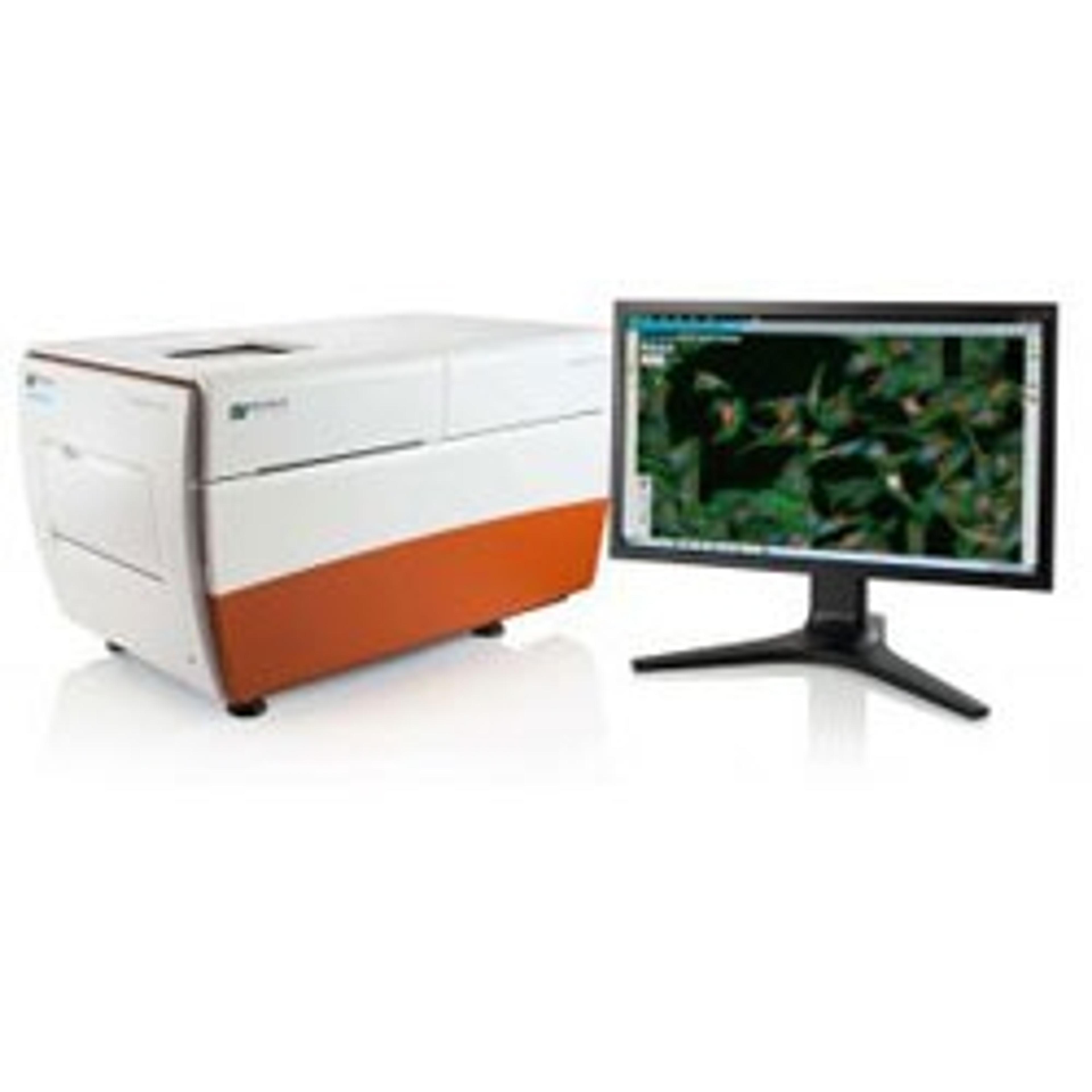
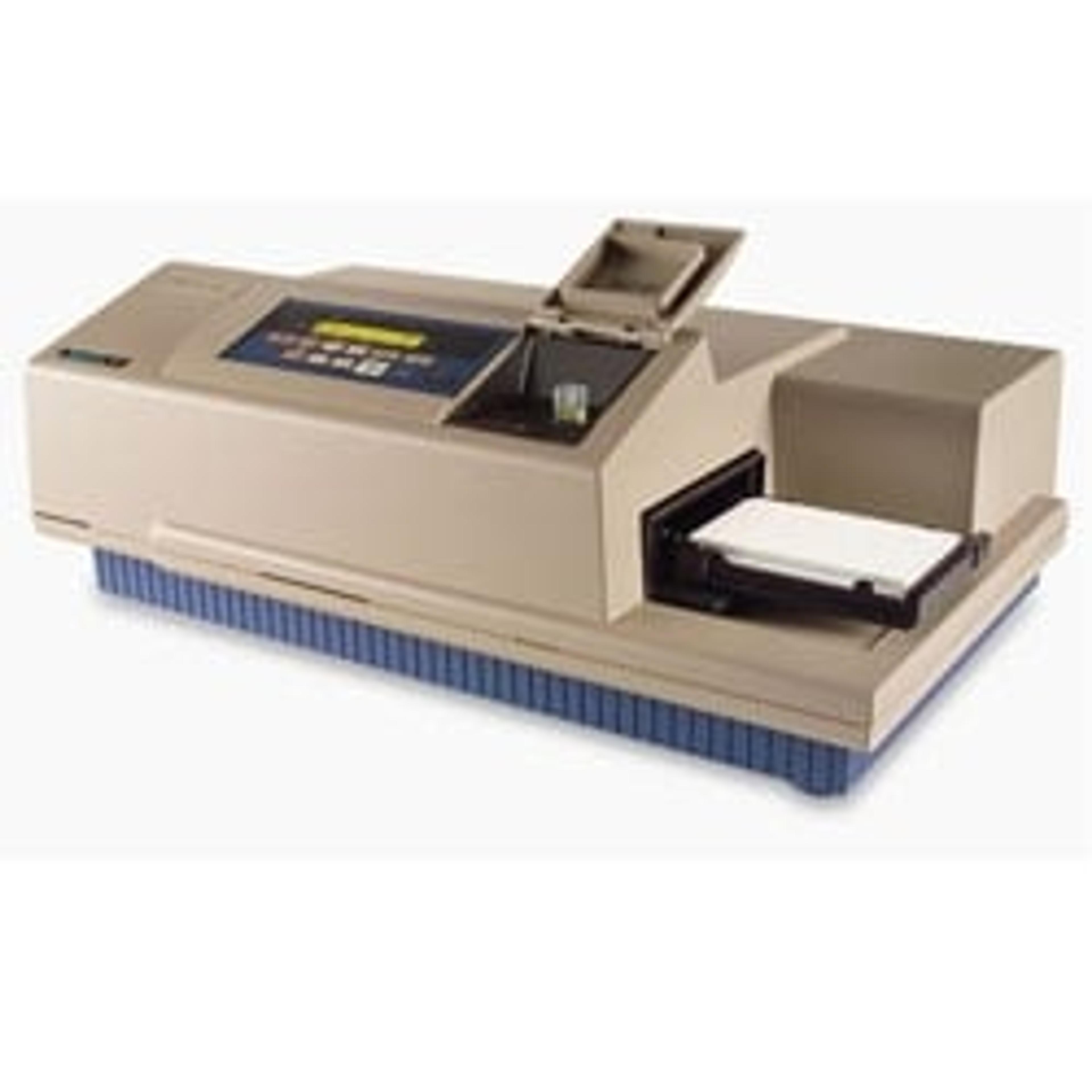
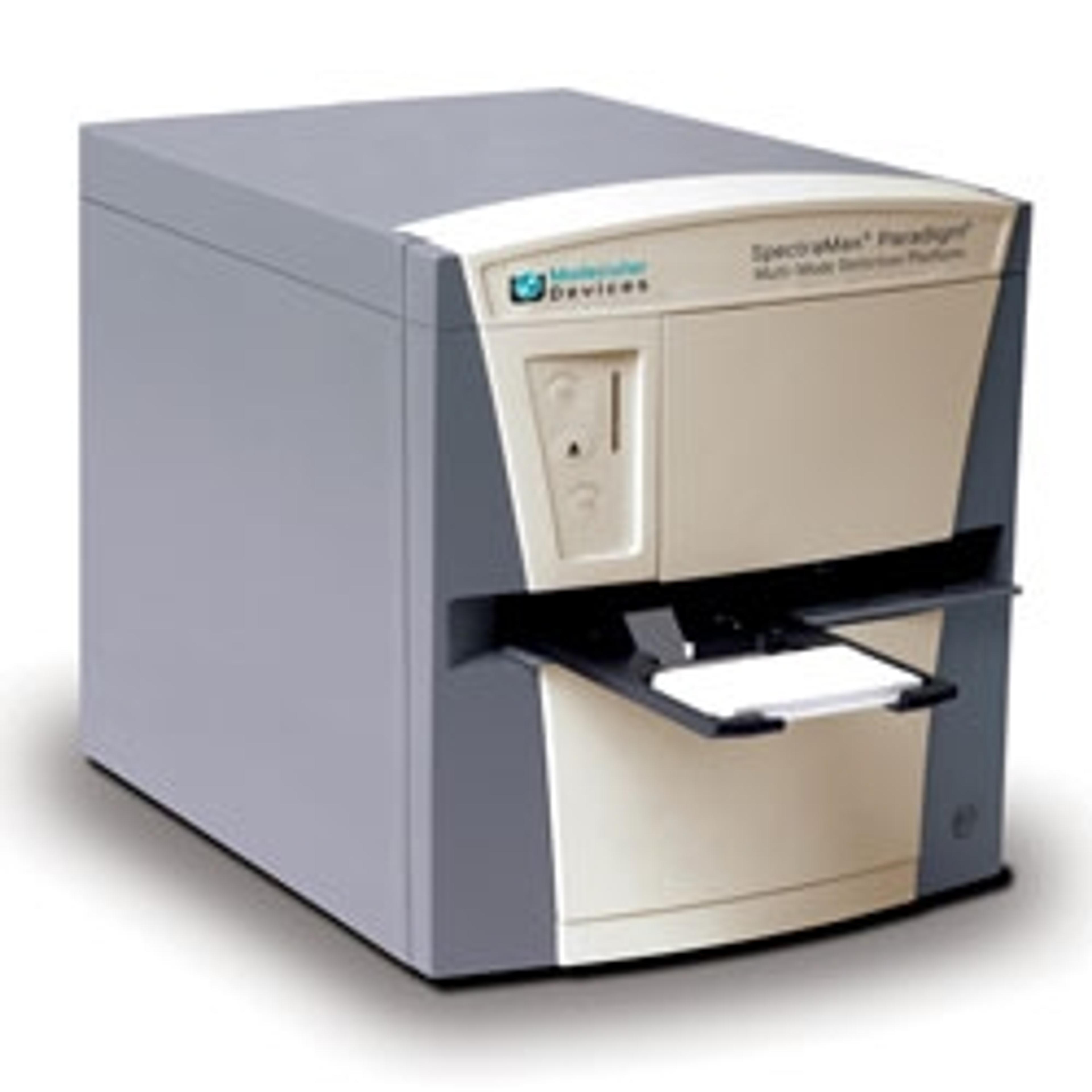
ImageXpress® Micro Confocal High-Content Imaging System
Explore the new dimension with the confocal system for your complex biology.





Receive your quote directly from the manufacturer.
The ImageXpress Micro Confocal system combines the ability to capture high-quality images without sacrificing throughput, reliability, or flexibility. Quickly and easily switch between confocal and widefield imaging modes to satisfy the specific throughput and sensitivity needs for your assay. Combined with MetaXpress® High-Content Image Acquisition and Analysis Software, the ImageXpress Micro Confocal system provides you with a complete multi-dimensional, high-throughput screening solution to help you discover your next landmark scientific breakthrough.For researchers looking to expand their laboratory’s capabilities, the ImageXpress Micro Confocal system leverages large field-of-view optics to map macro structures with minimal tiling. In addition, querying of large cell populations is accelerated, speeding up the characterization of highly heterogeneous samples or identification of rare sub-populations.
Key Benefits:
- Match acquisition mode to your assay needs with multiple confocal geometries and widefield mode
- Capture publication-quailty images without sacrificing throughput using our proprietary AgileOptix(TM) technology
- Acquire statistically relevant data faster with large field of view and > 3log dynamic range
- Expand your research capabilites with available options
The ImageXpress® Micro Confocal system represents the culmination of four generations of imaging expertise. Built on over 30 years of cell-based imaging experience, the ImageXpress Micro Confocal system is engineered for performance to reliably accelerate the pace of your research

Cellular imaging insights
The goal of many researchers is to elucidate the complex cellular pathways within 2D and 3D cellular structures in response to chemical stimulation or stress. The capacity to which scientists have access to and can adopt emerging cellular imaging and analysis technology fuels their level of understanding. In this eBook, Molecular Devices provides an overview of the new tools that can be used to better understand complex imaging models.

High-content phenotypic profiling using the Cell Painting assay
High-content phenotypic profiling is increasingly popular in research areas that span from gene function studies, drug discovery, and toxicology. The Cell Painting assay, often used in phenotypic profiling, uses up to six fluorescent dyes to label and visualize a variety of subcellular structures at the single cell level. The aim of this assay is to visualize as much of the cell as possible in order to construct a representative image of cell state. Molecular Devices shows how to simplify your Cell Painting assay workflow using the ImageXpress Micro Confocal system.
Key partnership increases access to organoid models
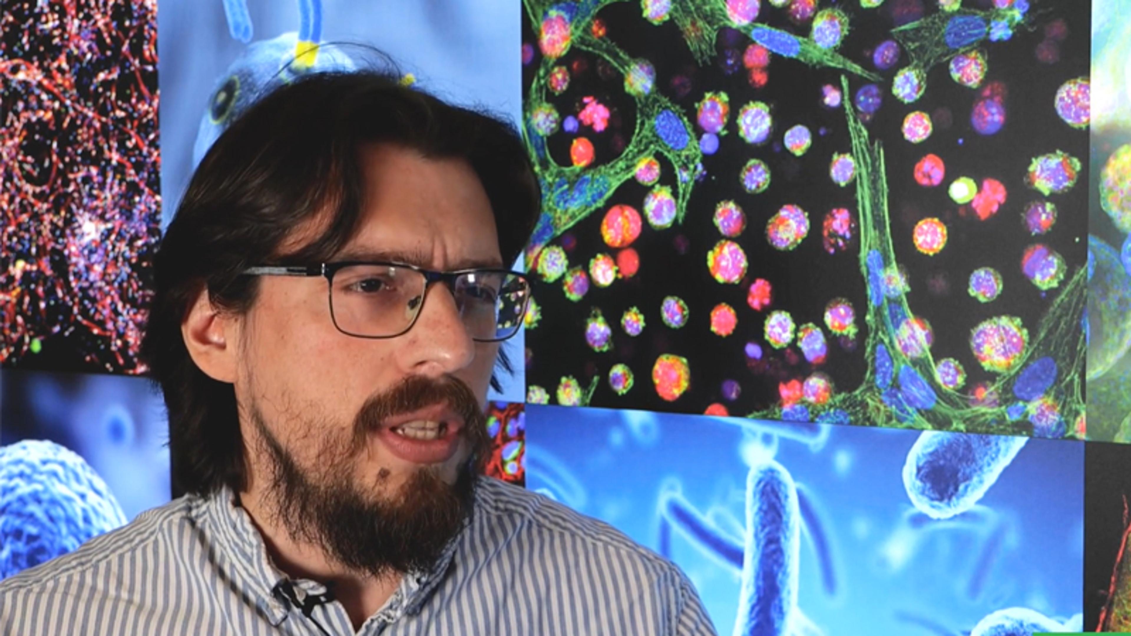
Patient-derived organoids address the urgent need for more predictive in vitro models for biopharmaceutical applications. However, challenges around standardization and scale up remain. Dr. Francisco Morales-Rodriguez shares insight into the partnership between HUB Organoids and Molecular Devices, which aims to increase the access of organoids for both academia and industry. Here, he highlights the integral role of the ImageXpress® Micro Confocal High-Content Imaging System and its suite of software solutions in providing high image quality, high throughput, and flexibility that HUB Organoids requires.
This video was filmed at SLAS Europe 2024.