Toward Quantitative Nanomechanical Measurements on Live Cells with PeakForce QNM
18 Dec 2013This application note reviews recent progress in mapping the properties of soft samples such as cells and gels with force volume and PeakForce QNM and the use of the newest NanoScopeR and NanoScope Analysis features to collect and analyze the data from these techniques.
Related products
Request Quote for All Products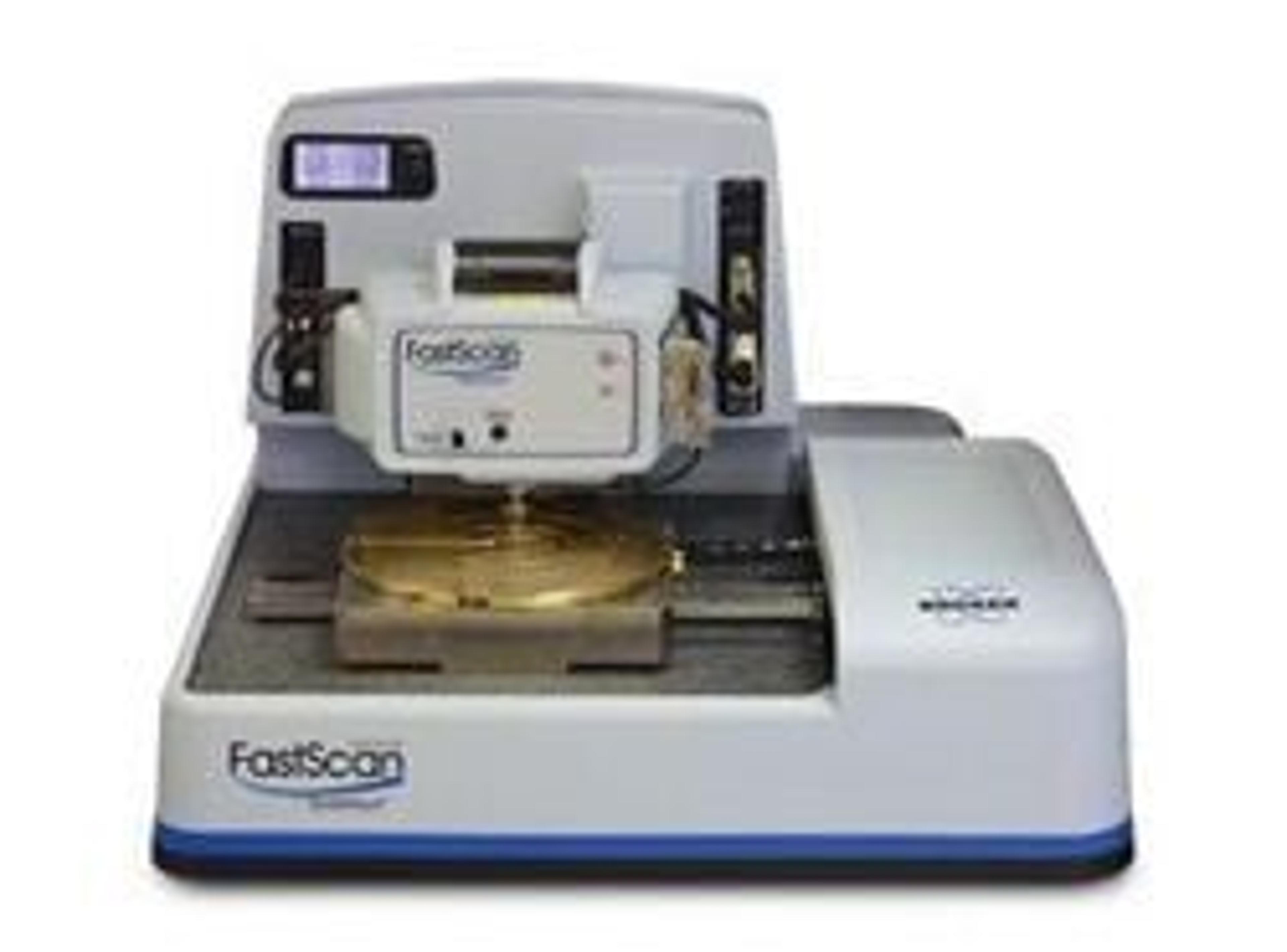
Dimension FastScan™
Bruker Nano Surfaces and MetrologyWorld's Ultimate AFM The new benchmark for speed with highest resolution and performanceThe Dimension FastScan™ delivers, for the first time, extreme imaging speed without loss of resolution, loss of force control, added complexity, or additional operating costs.This tip-scanning system provides measurements on both large and small size samples in air or fluids. With the FastScan you can achieve immediate atomic force microscopy images with the expected high resolution of a high-performance AFM, all in a single system. Whether surveying a sample scanning at >125Hz to find the region of interest, or scanning for detail at 1-second per image frame in air or fluids, the Dimension FastScan will redefine your AFM experience.Dimension FastScan™ Features: Work hundreds of times faster with fast scanning rates up to frames per second, automated laser and detector alignment, comprehensive work flow and smart engaging Built-in measurement automation software in conjunction with higher speed ScanAsyst® provide exceptional measurement confidence and repeatability Precise force control at the tip renders high resolution and long tip-life Low-noise, temperature-compensated sensors in the scanners maintain sub-nanometer noise levels Closed-loop Icon and FastScan scanners keep vertical noise below 30pm and 40pm, respectively, as well as high accuracy with ultra-low drift Sample from subnanometer to hundreds of nanometers in height without loss of resolution
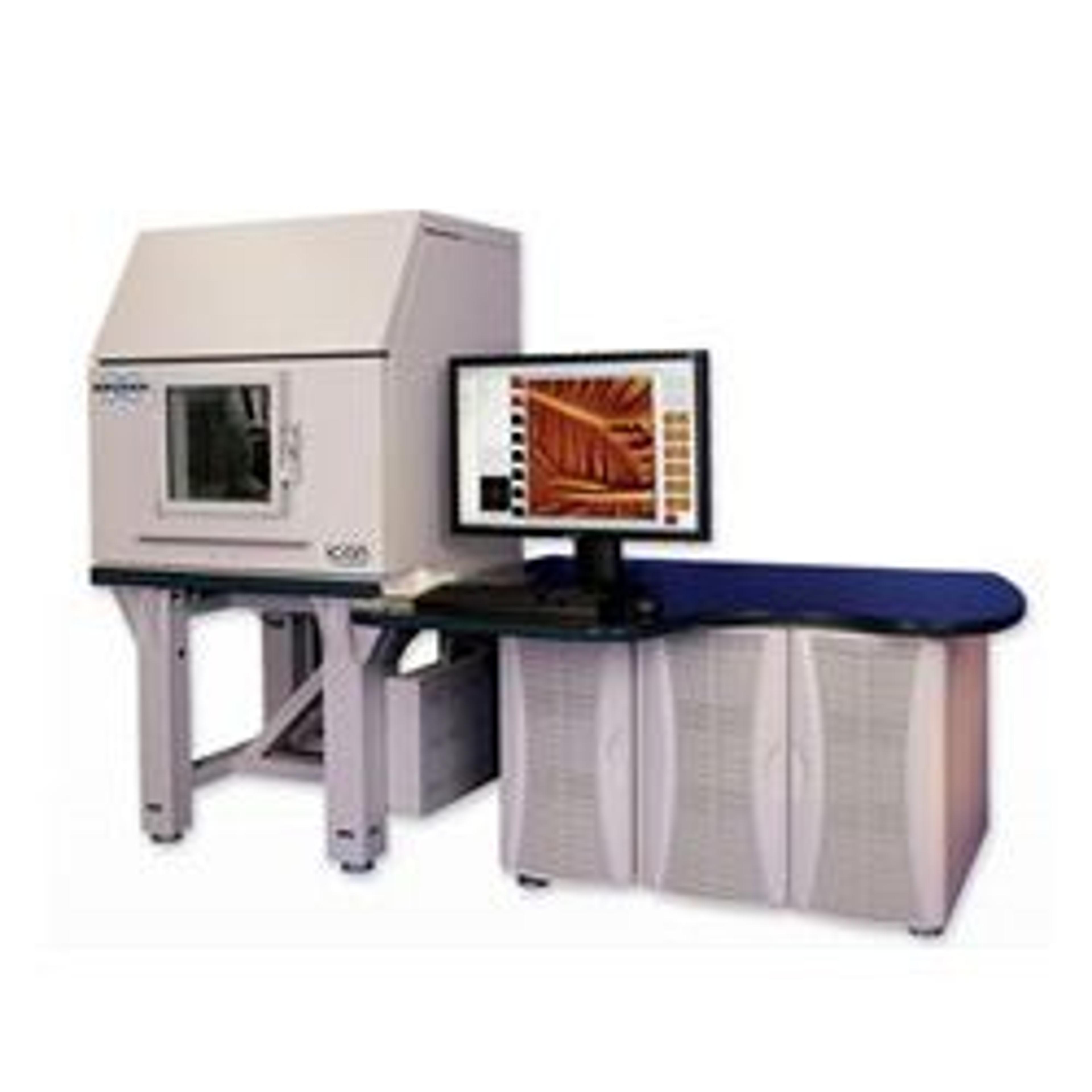
Dimension Icon Atomic Force Microscope
Bruker Nano Surfaces and MetrologyBringing new levels of performance, functionality, and AFM accessibility to nanoscale researchers. The Dimension Icon® Atomic Force Microscope brings new levels of performance, functionality, and AFM accessibility to nanoscale researchers in science and industry. The culmination of decades of large-sample AFM technology, the system has been designed from top to bottom to deliver revolutionary low drift and low noise that allows users to achieve artifact-free images in minutes instead of hours.The Icon is also equipped with proprietary ScanAsyst® automatic image optimization technology, which enables easier, faster, and more consistent results, regardless of user skill level. The Icon’s uncommon ease of use, ultimate performance, exceptional productivity, and superior versatility make it an ideal choice for practically every AFM application.Dimension Icon Atomic Force Microscope Features: Proprietary sensor design achieves closed-loop performance with open-loop noise levels for previously unseen resolution on large-sample AFMs Significantly reduced noise floor enables imaging at atomic level in contact mode, with less than 30pm in TappingMode™ Drift rates less than 200pm per minute render distortion-free images Integrated feedback alignment tools deliver quick and optimized probe positioning High-resolution camera and X-Y positioning permit faster, more efficient sample navigation ScanAsyst® Imaging and NanoScope® software with default experiment modes distill decades of knowledge into preconfigured settings Wide-open access to tip and sample accommodates a large variety of standard and customized experiments Instrument and software designed to take full advantage of all current and future Bruker AFM modes and techniques Custom user-programmable scripts offer semi-automated measurement and analysis