ResourceLife Sciences
Qualitative and quantitative evaluation of the tissue microenvironment by high-resolution 17-plex immunofluorescence reveals distinct populations
14 Jan 2022In this application note, Indica Labs highlights how the use of Orion imaging combined with HALO image analysis provides a powerful and intuitive workflow for visualization and quantification of distinct microenvironment populations.
Related products
Request Quote for All Products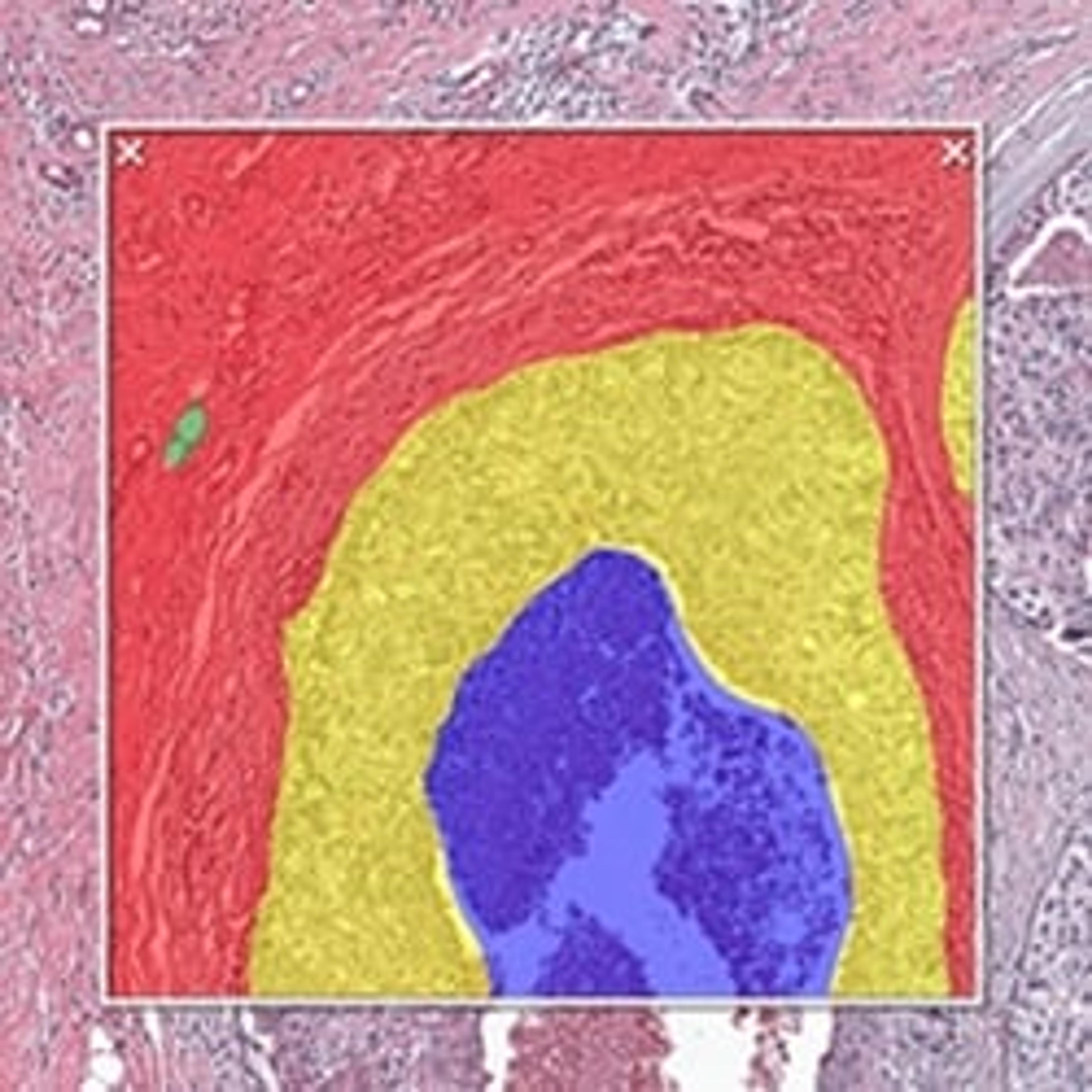
HALO® Image Analysis Platform
Indica LabsHALO provides easy-to-use, spatially-resolved cell and area-based image analysis for digital pathology and whole tissue images.
Tags
ImmunologyImmunological techniques measure and characterize immune responses. Immunology kits and analysis systems often use techniques such as ELISA, radioimmunoassay (RIA) and immunodiffusion assays, Immunohistochemistry, and flow cytometry. Immunologists use equipment such as flow Cytometers, plate readers, plate washers and fluorescent microscopes.Cellular PathologyCellular Pathology deals with the microscopic analysis of tissue samples and cells. Sample preparation and processing includes fixation, staining, sectioning and slide mounting, using equipment such microtomes and cryostats. In choosing immunohistochemistry and immunocytochemistry kits, consider chromogens, staining method, antibodies, microscopes and imaging.Light MicroscopyLight microscopes or optical microscopes are used to visualize microscale objects under magnification, including cells, clinical specimens and materials. Lab equipment for light microscopy includes confocal microscopes, fluorescence microscopes, zoom and stereo microscopes. Microscope slides and imaging reagents are available for visualizing samples, as well as various microscope stages and incubators for large or temperature-sensitive samples. Find the best light microscopes in our peer-reviewed product directory: compare products, check customer reviews and receive pricing direct from manufacturers.ImmunofluorescenceDigital PathologyDigital pathology involves the use of digital imaging and computational tools to analyze pathology slides. It is transforming diagnostics, research, and education by offering enhanced image storage and analysis. Explore digital pathology solutions in our peer-reviewed product directory; compare products, check reviews, and get pricing directly from manufacturers.MicroscopyMicroscopy is a technique used to observe small objects in detail, from cells to materials, using light or electron microscopes. It enables researchers to examine structures with high resolution, aiding in fields such as biology, medicine, and materials science. With advanced microscopy techniques, scientists can gain insights into cellular processes, tissue structures, and material properties. Explore the best microscopy solutions in our peer-reviewed product directory, compare products, read customer reviews, and get pricing directly from manufacturers.TumorsTumor research focuses on understanding abnormal cell growth that leads to cancer. Identifying biomarkers, studying tumor microenvironments, and developing targeted therapies are critical for advancing cancer treatment. Early detection and personalized treatment options are key to improving outcomes for patients. Browse our peer-reviewed product directory to explore tools for tumor research, diagnostics, and cancer therapies; compare products, read customer reviews, and get pricing directly from manufacturers.MultiplexingMultiplexing refers to the ability to measure multiple targets or analytes simultaneously in a single experiment. This technique is valuable for high-throughput screening, diagnostics, and complex assays, as it increases efficiency and data quality. Browse our peer-reviewed product directory to find the best multiplexing products, compare tools, check reviews, and get pricing directly from manufacturers.