HALO AI
Neural network driven tissue and cell classification for research & routine pathology
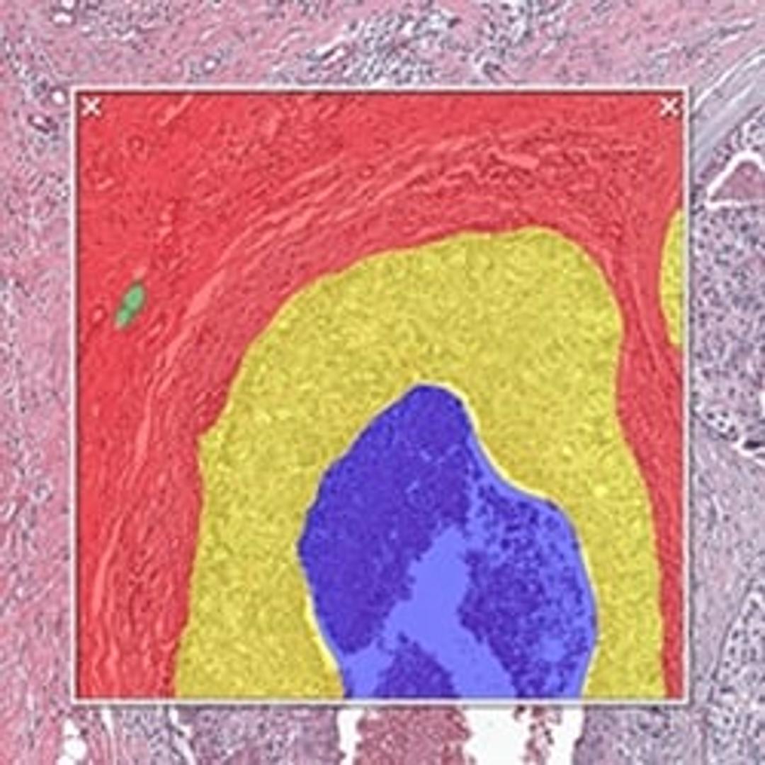
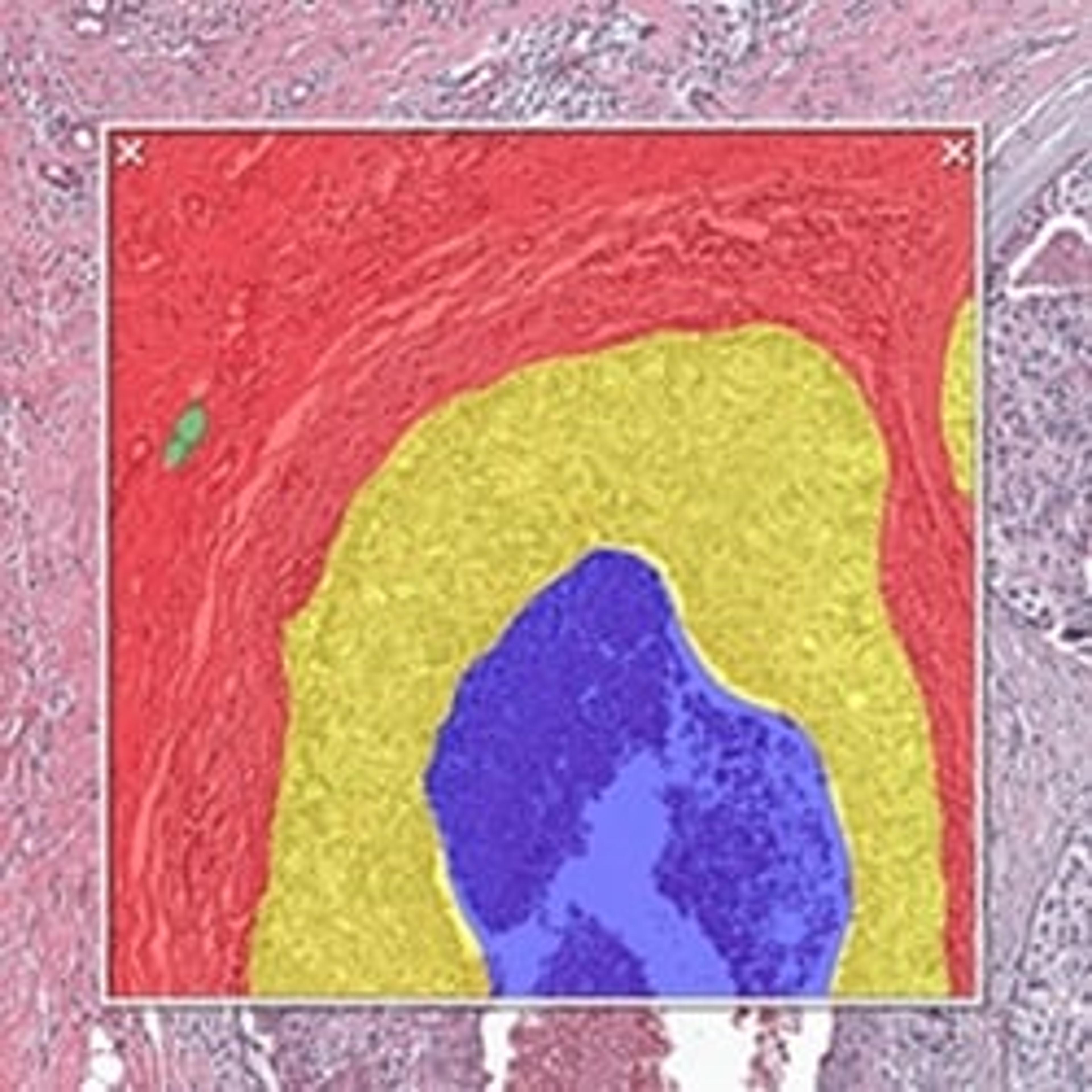
HALO AI classification of breast cancer H&E




HALO AI is a collection of train-by-example classification and segmentation tools underpinned by advanced deep learning neural network algorithms. HALO AI classifiers can be trained to quantify tissue classes, to segment tissue classes for analysis with other HALO image analysis modules, to find rare events or cells in tissues, and to categorize cell populations into specific phenotypes.
SIMPLE & INTUITIVE WORKFLOW
HALO AI is fully integrated with the intuitive, easy-to-use HALO and HALO Link viewers and employs a simple three-step workflow. After defining what tissue classes or cell phenotypes you would like to segment, you train the neural network by drawing annotations – no computer programming or AI knowledge required. Trained classifiers can be applied to segment tissue and cells on any whole slide image or region of interest.
POWERFUL TISSUE SEGMENTATION
HALO AI now includes the option of three powerful neural networks – VGG, DenseNet and MiniNet. VGG, a well-known and more traditional network, was used to build the Indica Labs submission in the CAMELYON17 challenge and was the first neural network integrated with HALO AI. DenseNet is a more modern network capable of creating more robust classifiers at higher resolution compared to VGG. MiniNet, a custom network developed at Indica Labs, is more shallow than VGG or DenseNet, but can produce a solution quickly with limited training data and is therefore useful for testing new AI applications.
EXCEPTIONAL CELL CLASSIFICATION
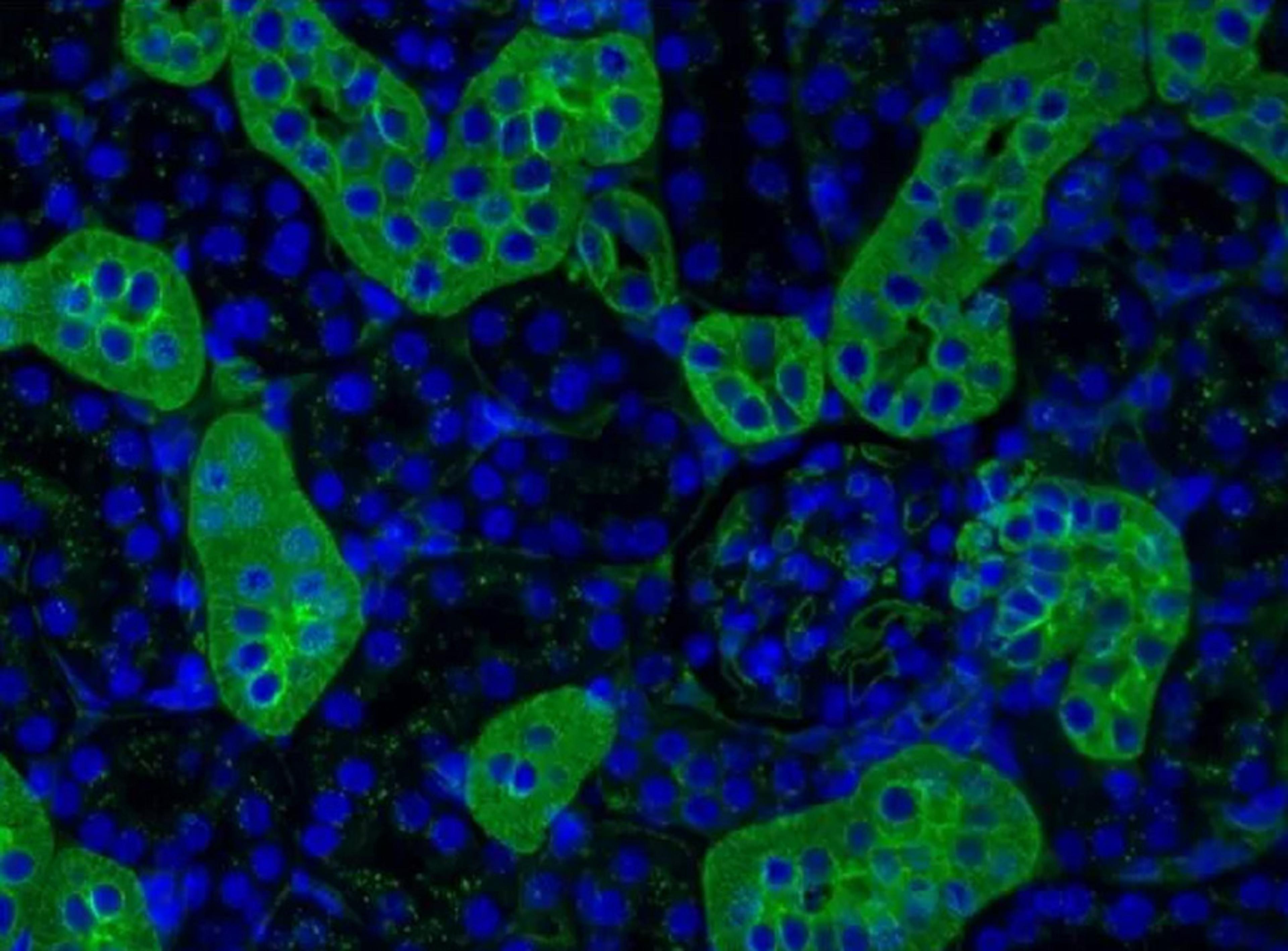
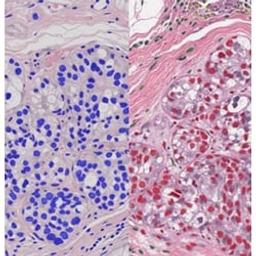
Segment nuclei with the new Segmentation classifier. Utilize HALO AI’s pretrained networks for H&E, single IHC, or DAPI stained images for an out of the box solution. Or train your own nuclei segmentation network for a specific application (unique tissue or advanced staining protocols). Once nuclei are segmented, take it a step further using the Nuclei Phenotyper classifier to automatically assign cells into user defined phenotypes with a few quick training examples.

Introduction of a robust workflow for the whole-slide acquisition and co-registration of multiplex immunofluorescence tissue images
In this application note, explore how Indica Labs designed and tested an 8-plex immunophenotyping panel using the InSituPlex® approach to detect and classify T cells, macrophages, and tumor cell in non-small cell lung cancer and colorectal cancer FFPE tissue.


Xyall and Indica Labs forge global collaboration
The partnership is designed to bridge the gap between histopathology and molecular pathology