ResourceLife Sciences
Monitor mitochondria dynamics and phenotype with high-content imaging
8 Dec 2021Mitochondria are the main energy source for cells and play a key role in regulating cellular metabolism. In this application note, Molecular Devices describes phenotypic assays for mitochondria phenotypes and structural re-arrangements that can be used for studies of mitochondria dynamics in cell-based assays.
Related products
Request Quote for All Products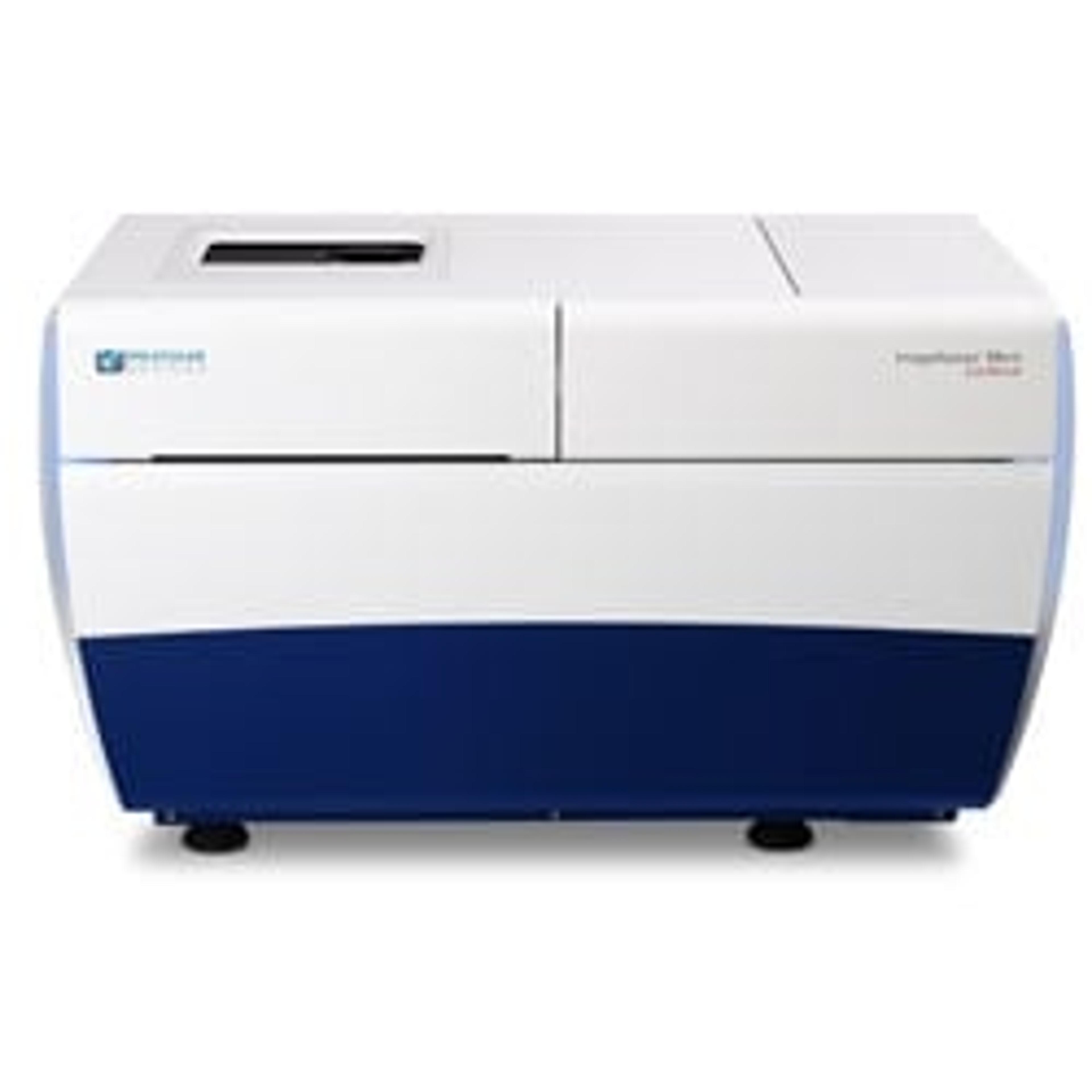
ImageXpress® Micro Confocal High-Content Imaging System
Molecular Devices®Explore the new dimension with the confocal system for your complex biology.
Links
Tags
In Vivo Imaging Systems<i>In vivo</i> imaging systems, including pre-clinical imaging systems and medical imaging systems are used to non-invasively visualize and capture images of live animals and plants. Monitor the natural processes or diseases of your subjects using small-animal pre-clinical imaging systems, including single photon positron emission tomography (SPECT), positron emission tomography (PET), computed tomography (micro-CT), magnetic resonance imaging (MRI), X-ray radiography, ultrasound, fluorescence and bioluminescence imagers. Multimodal systems and software solutions are also available for correlative analysis of organ, tissue, cell, or molecular-level processes. Find the best in vivo imaging products in our peer-reviewed product directory: compare products, check customer reviews and receive pricing direct from manufacturers.MitochondriaHigh Content ImagingHigh content imaging is a method combining two or more fluorescent microscopy experiments to identify substances that alter a cell’s phenotype in a desired manner. The process is adapted to multi-well plates and both the image acquisition and analysis are automated.Phenotypic ScreeningPhenotypic screening assesses cellular responses to compounds, enabling drug discovery and target identification. This technique is pivotal in finding effective treatments for complex diseases. Discover phenotypic screening platforms and tools with peer-reviewed comparisons and pricing in our directory.