ResourceLife Sciences
Increase Sensitivity in No-Wash Assays Using Confocal Imaging
15 Feb 2019Screening of hybridoma supernatants for antibodies directed against cell-surface antigens is an important step in the discovery and development of antibodies for vaccines or therapeutic use. The sensitivity of these assays is increased by taking advantage of the optical properties of confocal imaging such as utilizing the narrow depth of field to acquire high signal-to-background, crisp images with no out of focus light creating image blur.
Related products
Request Quote for All Products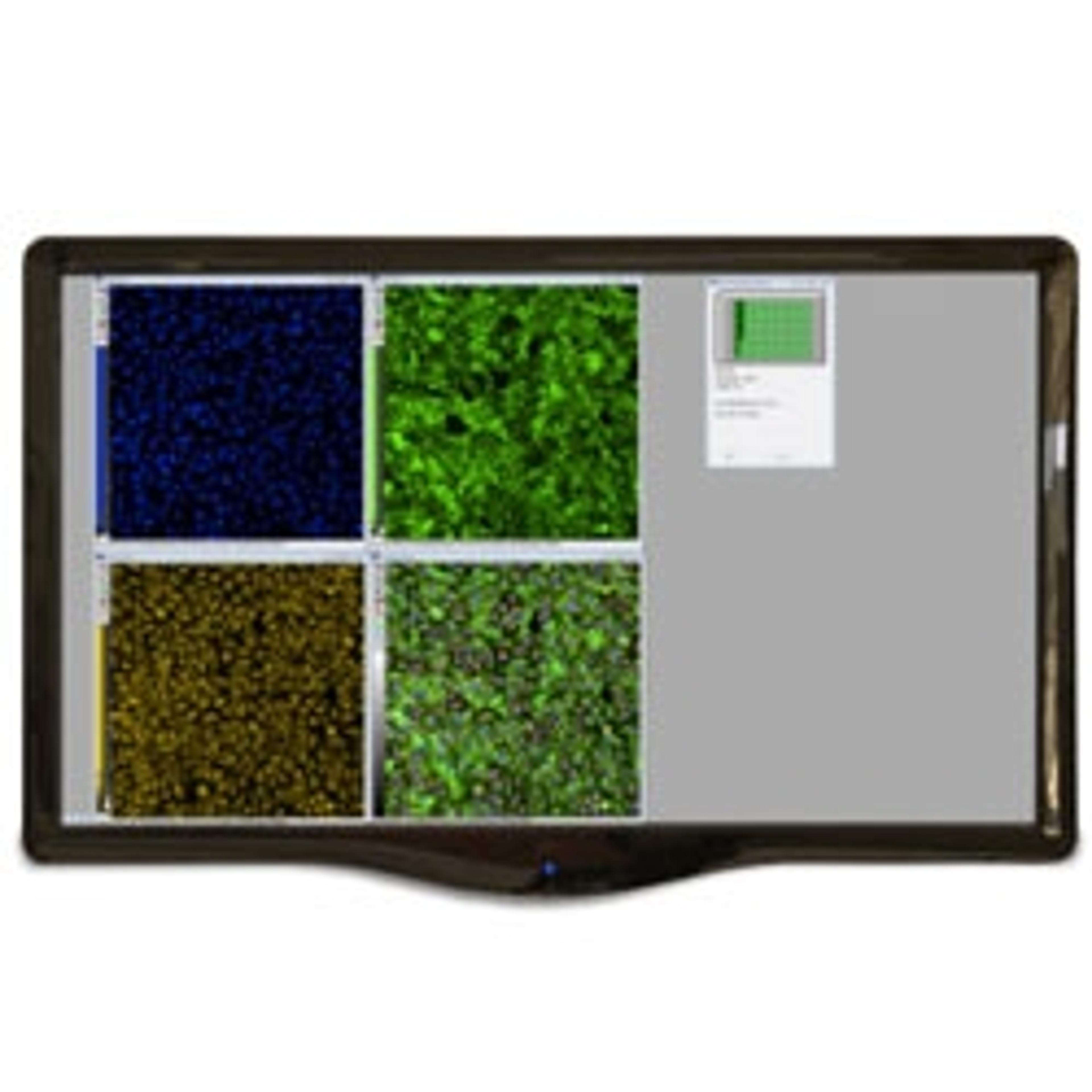
MetaXpress High-Content Image Acquisition and Analysis Software
Molecular Devices®High-content image analysis software featuring time lapse analysis
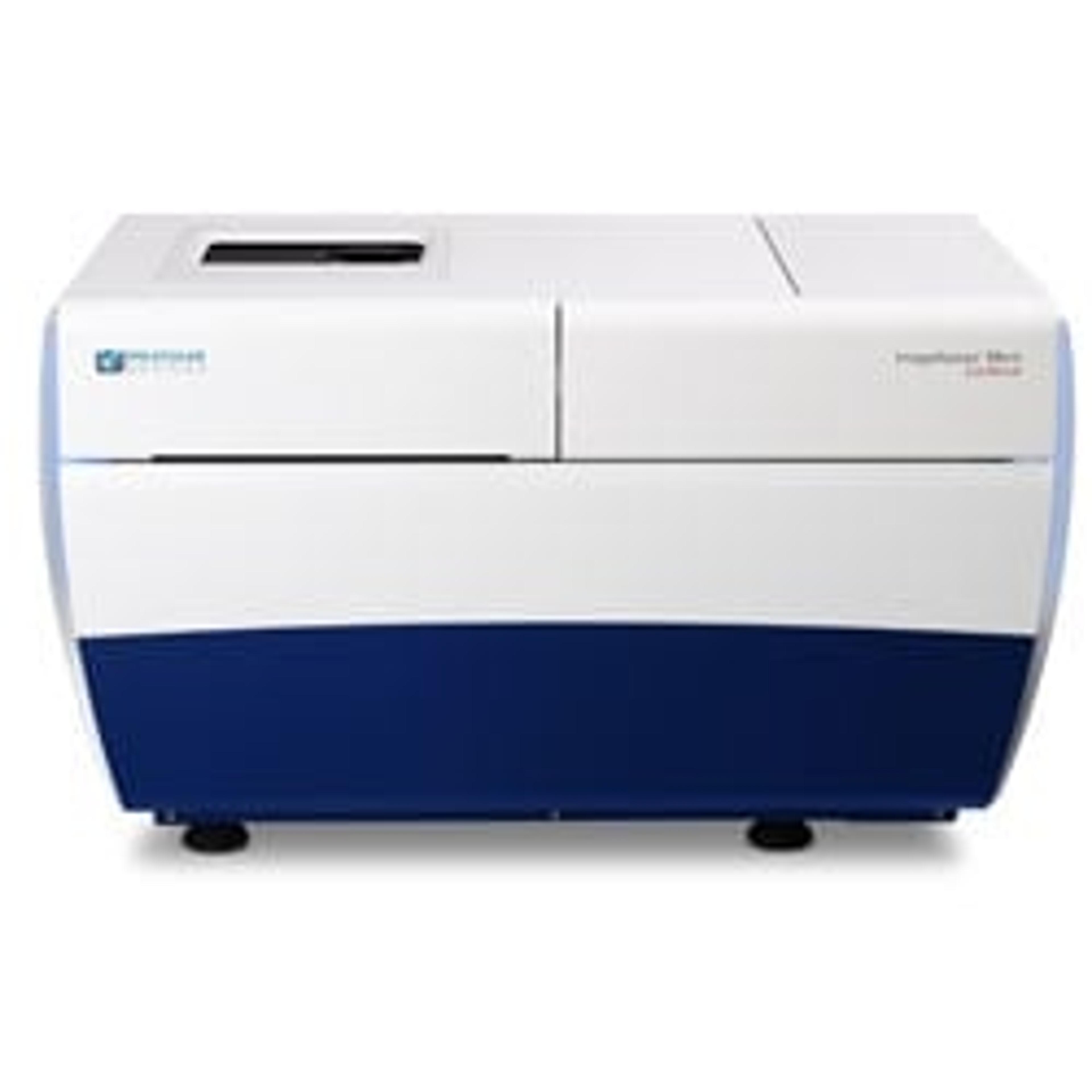
ImageXpress® Micro Confocal High-Content Imaging System
Molecular Devices®Explore the new dimension with the confocal system for your complex biology.
Links
Tags
AntibodiesAntibodies are used in techniques such as confocal and fluorescence microscopy, flow cytometry, ELISA, ELISPOT, immunohistochemistry, western blotting and immunopreciptation. Select specific antigen reactivity, high specific affinity, low non-specific binding, monoclonal or polyclonal, primary or secondary antibodies and associated conjugates such as an enzyme or dye for visualization.High-Content ScreeningHigh-content screening (HCS), also known as high-content analysis (HCA), is a high-throughput technique used in drug discovery to identify substances that alter the phenotype of cells. HCS uses fluorescent microscopic imaging and automated image analysis to investigate cellular events such as apoptosis, cell viability, GPCR activation, oxide production, neurite outgrowth, and cell signaling. Find the best fluorescent labeling reagents, cellular assays, and high-content imaging systems in our peer-reviewed product directory: compare products, check customer reviews and receive pricing direct from manufacturers.Fluorescence Based AssayFluorescence based assays are widely used in life science research and high-throughput screening to measure a broad range of cellular activities.ConfocalHigh Content ImagingHigh content imaging is a method combining two or more fluorescent microscopy experiments to identify substances that alter a cell’s phenotype in a desired manner. The process is adapted to multi-well plates and both the image acquisition and analysis are automated.