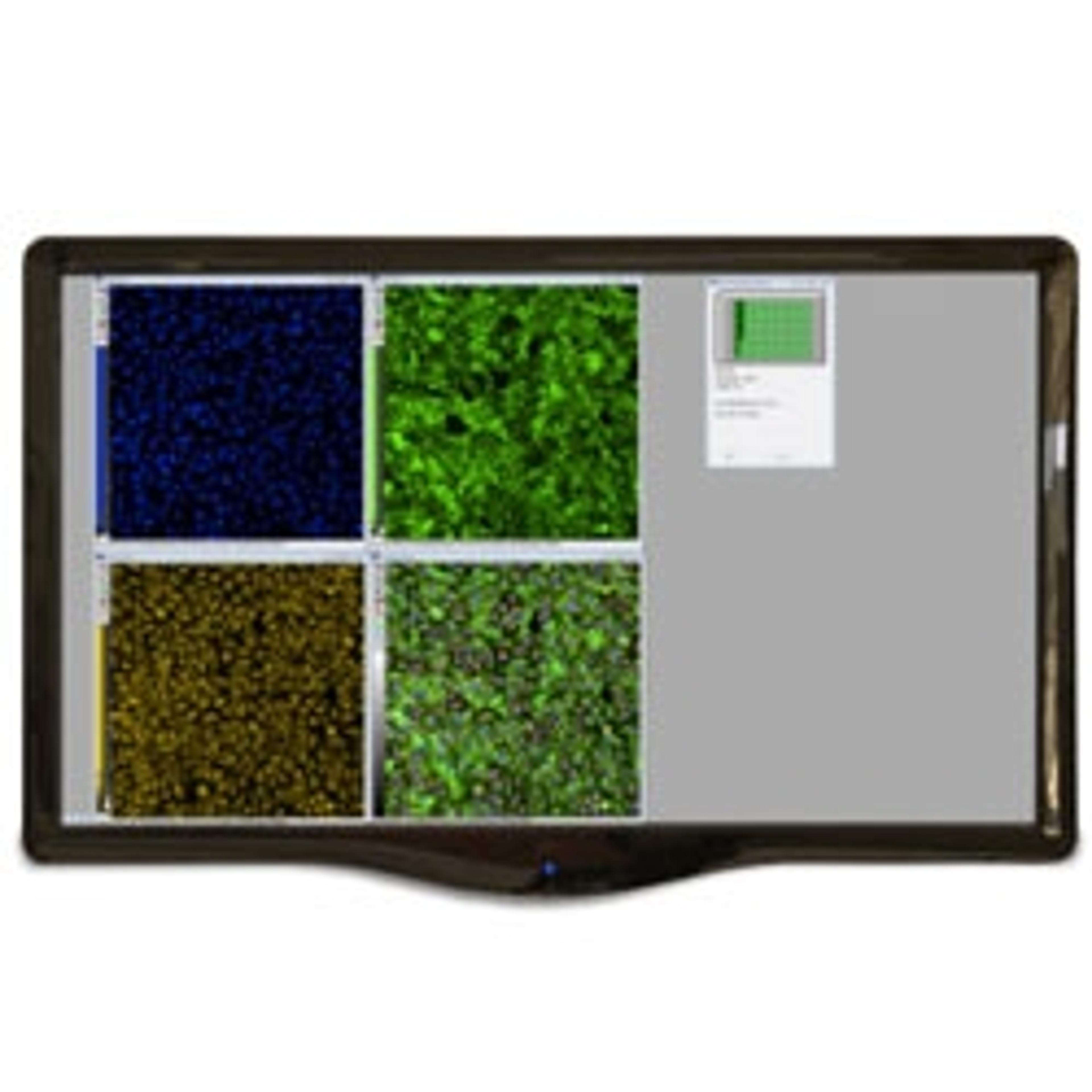
High-Content Assay for Morphological Characterization of 3D Neuronal Networks in A Microfluidic Platform
29 Mar 2019The focus of this application note is to develop a high-throughput 3D neurite outgrowth assay using iPSC-derived neurons developed in the microfluidic OrganoPlate® platform, with the goal of establishing 3D models for neurodegenerative diseases and neurotoxicology screens. The OrganoPlate® is a high-throughput platform that combines the most recent advances in 3D cell culture, Phaseguides™ and microfluidics. The OrganoPlate contains 96 tissue chips suitable for long-term culture of live cells, is amenable for screening purposes, and is compatible with standard laboratory equipment or automated systems, such as the ImageXpress® Micro Confocal High-Content Imaging System.
Related products
Request Quote for All Products
OrganoPlate® 3-lane
MIMETAS B.V.The OrganoPlate 3-lane features 40 independent culture cells, each supporting adjacent in-gel culture and 2 perfusion channels, that can contain perfused tubular tissues. The 3-lane models supports apical and basal access to epithelial and endothelial tubules, allowing barrier integrity and transport assays. The lanes are 400 micron wide. Six wells of the 384-well-plate user interface connect to each microfluidic network in a 9x9 well grid, the central well provides viewing access to the microtissue.
MetaXpress High-Content Image Acquisition and Analysis Software
Molecular Devices®High-content image analysis software featuring time lapse analysis
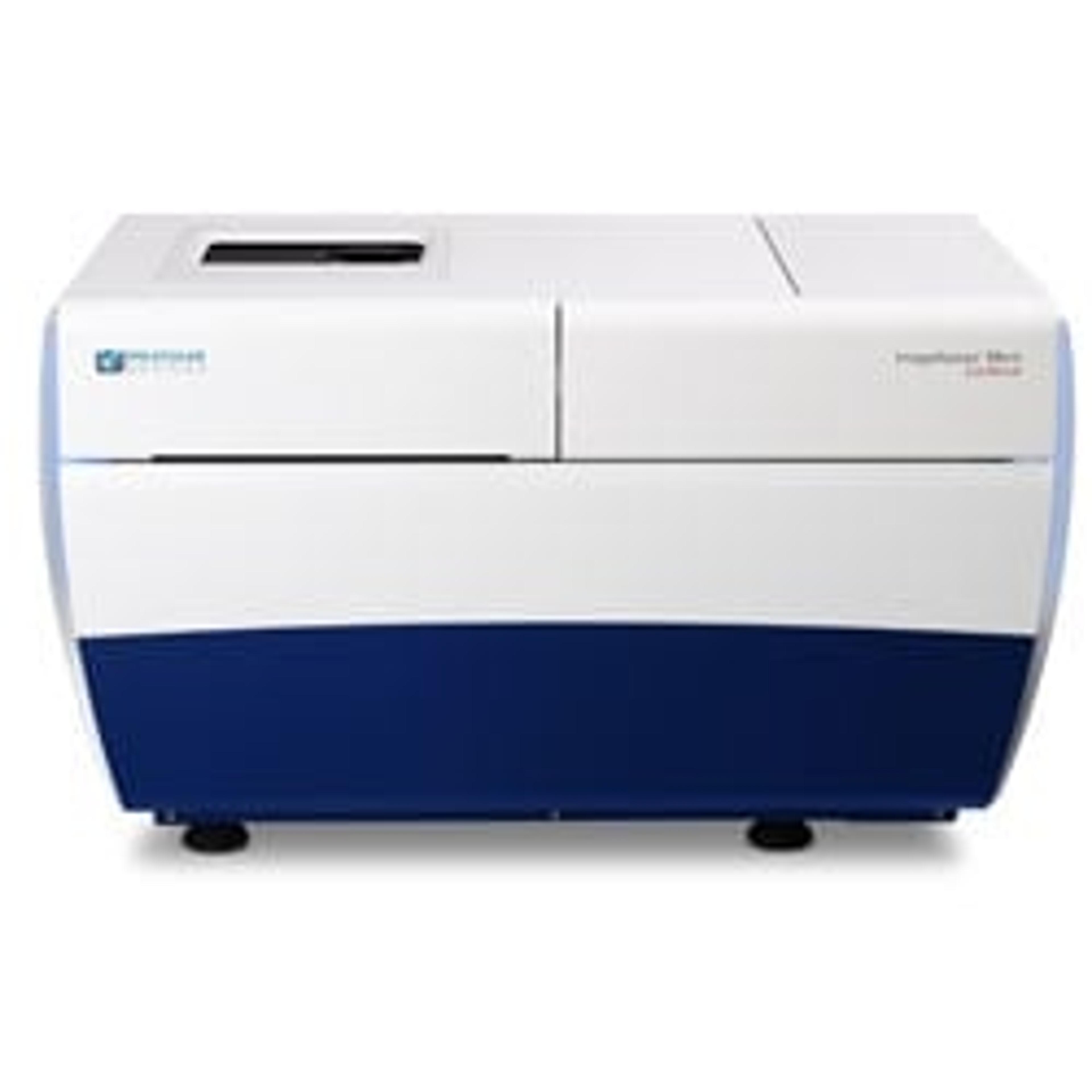
ImageXpress® Micro Confocal High-Content Imaging System
Molecular Devices®Explore the new dimension with the confocal system for your complex biology.