ResourceLife Sciences
Assessment of Drug Effects on Cardiomyocyte Physiology Using Human iPSC-Derived Cardiac Spheroids
9 Mar 2019Cell models are becoming more complex in order to better mimic in vivo microenvironments and provide greater predictivity for compound efficacy and toxicity. There is an increasing interest in exploring the use of three-dimensional (3D) spheroids for modelling tissue biology and toxicity assessment. Development of quantitative assays in higher throughput using 3D cultures is an active area of investigation. In this study, we have developed methods for the formation of 3D spheroids derived from human induced pluripotent stem cells (iPSC).
Related products
Request Quote for All Products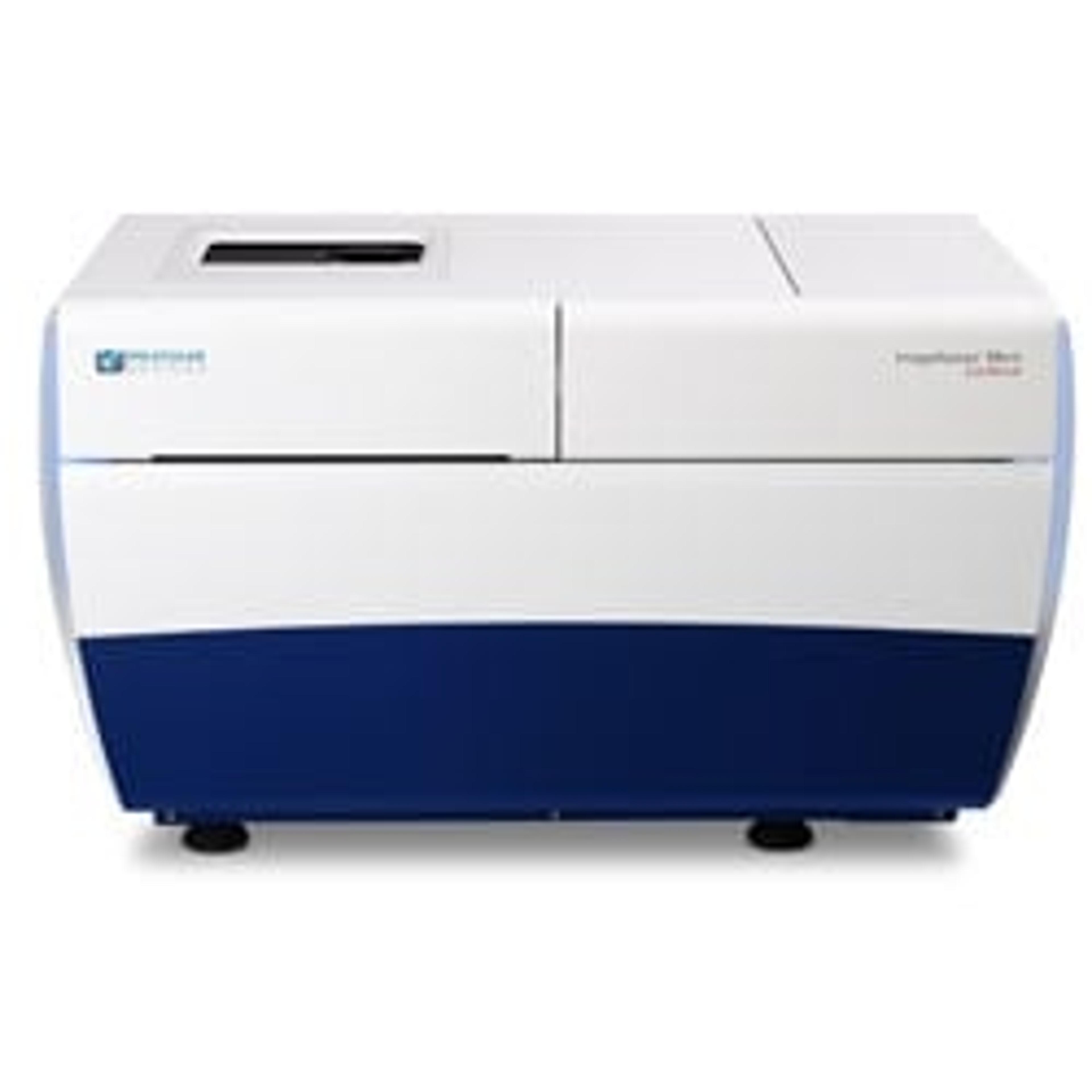
ImageXpress® Micro Confocal High-Content Imaging System
Molecular Devices®Explore the new dimension with the confocal system for your complex biology.
Links
Tags
Cell / Tissue CultureCell culture or tissue culture is used to study the biology of cells or tissues and to isolate cellular products in an environment which can be manipulated and well defined. Accurately control your culture environment with bioreactors or culture incubators, bind your cells to a surface or together with an extracellular matrix. Distinguish cell types with differential media or proliferate cells with certain characteristics using selective media. Enrich your media with supplements such as growth factors, sera and vitamins. Find the best cell and tissue culture products, kits and equipment in our peer-reviewed product directory: compare products, check customer reviews and receive pricing direct from manufacturers.High-Content ScreeningHigh-content screening (HCS), also known as high-content analysis (HCA), is a high-throughput technique used in drug discovery to identify substances that alter the phenotype of cells. HCS uses fluorescent microscopic imaging and automated image analysis to investigate cellular events such as apoptosis, cell viability, GPCR activation, oxide production, neurite outgrowth, and cell signaling. Find the best fluorescent labeling reagents, cellular assays, and high-content imaging systems in our peer-reviewed product directory: compare products, check customer reviews and receive pricing direct from manufacturers.FluorescenceThe emission of fluorescence occurs when a photon of energy is supplied to a fluorescent chemical compound by an external source, causing it to become excited. Fluorescence can be detected and measured for different purposes using microplate readers, fluorescence microscopes, fluorescence scanners, and flow cytometers.Human CardiomyocytesHigh ThroughputHigh throughput experiments allow the simultaneous processing of several samples. This parallelization reduces the cost per experiment and increases reproducibility and output volume of data.CardiomyocytesHigh Content ImagingHigh content imaging is a method combining two or more fluorescent microscopy experiments to identify substances that alter a cell’s phenotype in a desired manner. The process is adapted to multi-well plates and both the image acquisition and analysis are automated.SpheroidsSpheroids are clusters of cells that have been grown in 3D culture to be used as <i>in vitro</i> model systems. These 3D microtissues can be used for toxicology testing, DMPK studies and many other applications involving cell analysis.ScreeningUsing robotics, data processing and control software, liquid handling devices and sensitive detectors, screening allows a researcher to quickly conduct millions of chemical, genetic or pharmacological tests.hiPS Cell Lines