CellScape™ Platform
High plex proteomics instrument for spatial biology
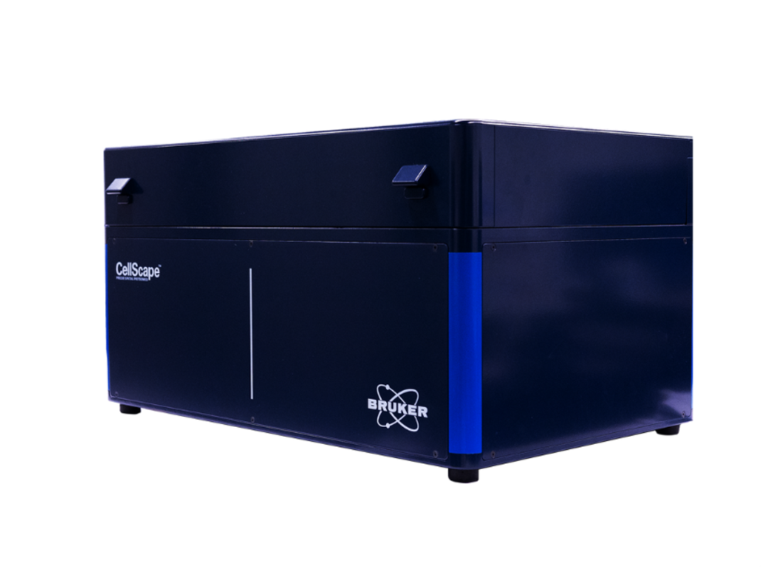
CellScape™ Platform


Receive your quote directly from the manufacturer.
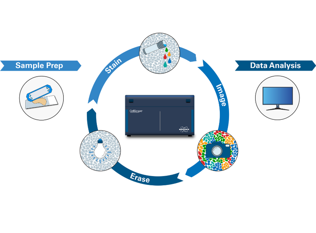
CellScape™ is the next generation in ChipCytometry™ instrumentation, advancing the cutting-edge platform for quantitative in situ spatial phenotyping. ChipCytometry delivers single-cell targeted spatial proteomics for complex whole-tissue analysis of the tumor microenvironment, as well as deep immune profiling for applications in immunology, neuroscience, and infectious disease. The new system builds on the existing core strengths of the original ChipCytometry instrument, the ZellScannerONE™, which has enabled spatial biology research on both tissue samples and cell suspensions since 2016.
CellScape, with its multiplexed fluidics integration, adds complete walk-away automation, improved optical performance, and massively increased throughput for whole slide imaging of millions of cells, while maintaining the key features of the core ChipCytometry technology, including high-plex phenotyping with single-cell resolution, high dynamic range imaging for detection and quantification of both high- and low-expressing targets, and compatibility with standard commercially available fluorescently labelled antibodies, requiring no proprietary antibody conjugation. This powerful combination of features will significantly improve researchers’ workflow, accelerate spatial biology discovery, and drive broader adoption of high-plex spatial omics for translational and clinical applications.
Brochures

Precise spatial proteomics with the CellScape Platform
Explore the CellScape™ platform from Bruker Spatial Biology – an end-to-end solution for highly multiplexed spatial proteomics and single-cell analysis.* With an advanced imaging system, streamlined fluidics for walk-away automation, and unprecedented flexibility in assay design, the CellScape platform accelerates biological research from discovery to translation.
*For research use only. Not for use in diagnostic procedures.
Expandable immunofluorescence assays with next-generation VistaPlex immune profiling kits
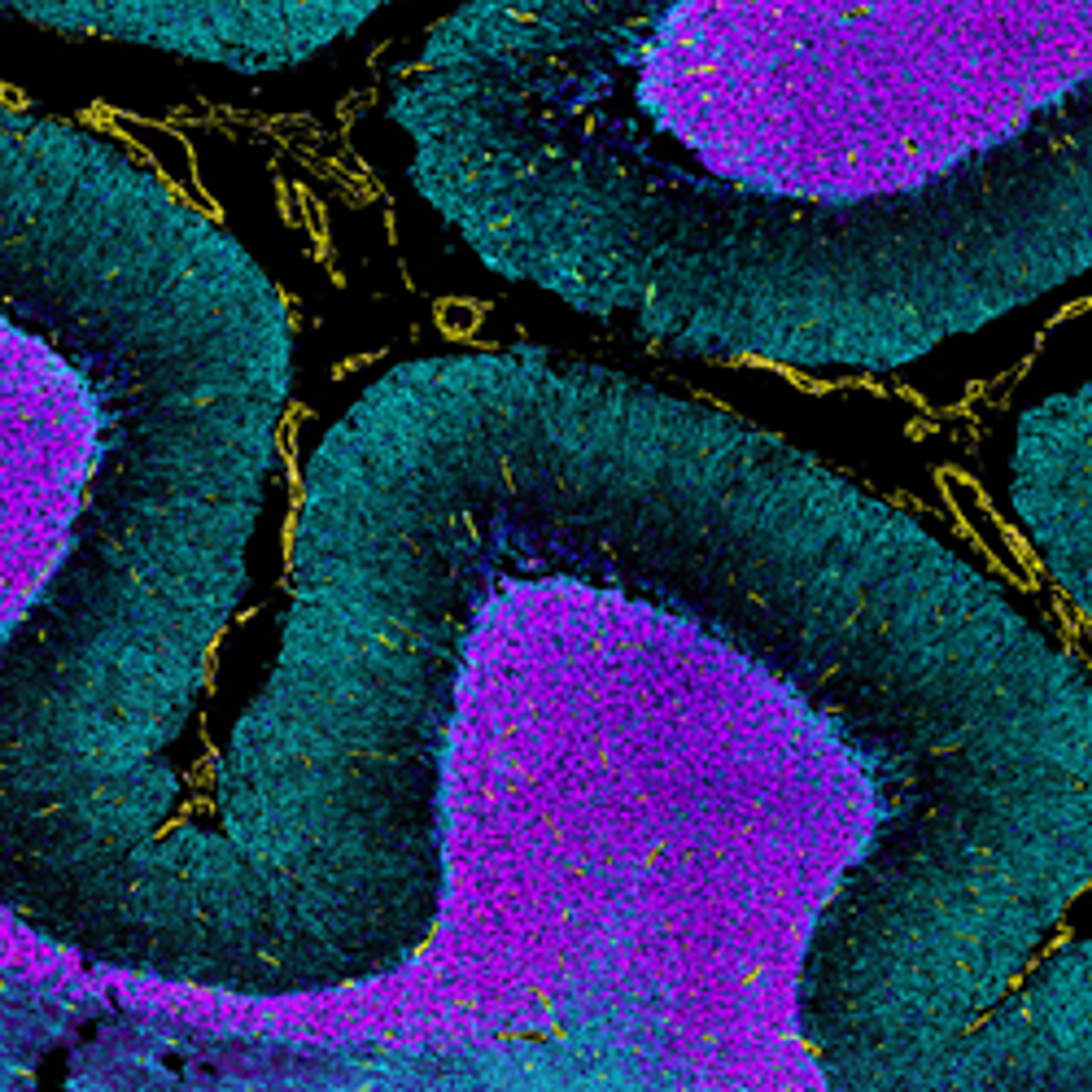
VistaPlex assay kits are modular, ready-to-use multiplex immunofluorescence (mIF) assays designed to deliver comprehensive cellular phenotyping and spatial tissue insights for advanced spatial biology applications. Enhanced by EpicIF technology, the CellScape Precise Spatial Proteomics platform now supports expanded multiplex dye compatibility, ensuring that next-generation VistaPlex kits integrate seamlessly with the EpicIF workflow. Data presented highlight the effectiveness of the Segmentation, Spatial Immune Profiling, and Architecture panels across a wide array of human FFPE tissues, revealing distinct biological insights achievable with individual kits or combined panel configurations.
VistaPlex immunofluorescence assay panels
VistaPlex Spatial Immune and Architecture Kits for spatial analysis on human formaldehyde-fixed, paraffin-embedded (FFPE) tissues enable the profiling of immune populations and tissue architecture using validated antibodies and optimized staining protocols. Canopy Biosciences details a study which compares the efficiency of different assay kits and highlights their ability to identify various cellular phenotypes across different tissue types and disease states. Further, Canopy Biosciences emphasizes the compatibility of VistaPlex kits with CellScape Whole-Slide Imaging Chambers, enhancing the reproducibility and utility of the assays in basic and translational research settings. Overall, VistaPlex kits offer a robust and cost-effective solution for spatial biology research, facilitating deep phenotyping of immune and cancer cells in diverse tissues.
Precise spatial multiplexing of protein biomarkers for immune profiling in tissue samples with ChipCytometry
Immunohistochemistry is the most widely used diagnostic technique in tissue pathology. However, IHC is associated with several limitations including the labeling of just a few markers per tissue section and limited quantification of cell populations. As a result of plex limitations, key insights about tumor biology are missed, which could be important for advancing our understanding of tumor biology and ultimately improving patient outcomes. ChipCytometry™ is a novel image-based platform for precise spatial multiplexing that addresses these challenges by combining iterative immuno-fluorescent staining with high dynamic range imaging to facilitate quantitative phenotyping with single-cell resolution.
In this research poster, Canopy Biosciences demonstrates how standard flow cytometry standard (FCS) files are generated from multichannel OME-TIFF images, enabling the identification of cellular phenotypes via flow cytometry-like hierarchical gating. Quantification of results reveals precise expression levels for each marker in the assay in each individual cell in the sample, while maintaining spatial information about each cell.
Bruker Spatial Biology to announce groundbreaking advances at AGBT 2025
Advancements to include CosMx® Whole Transcriptome Panel; enhanced technology engine to power CellScape™ for spatial proteomics; expansion to 1000-plex protein assay on GeoMx® DSP; and launch of PaintScape™, a revolutionary platform enabling direct visualization of the 3D genome
Product Overview

Links
Featured products
Request Quote for All Products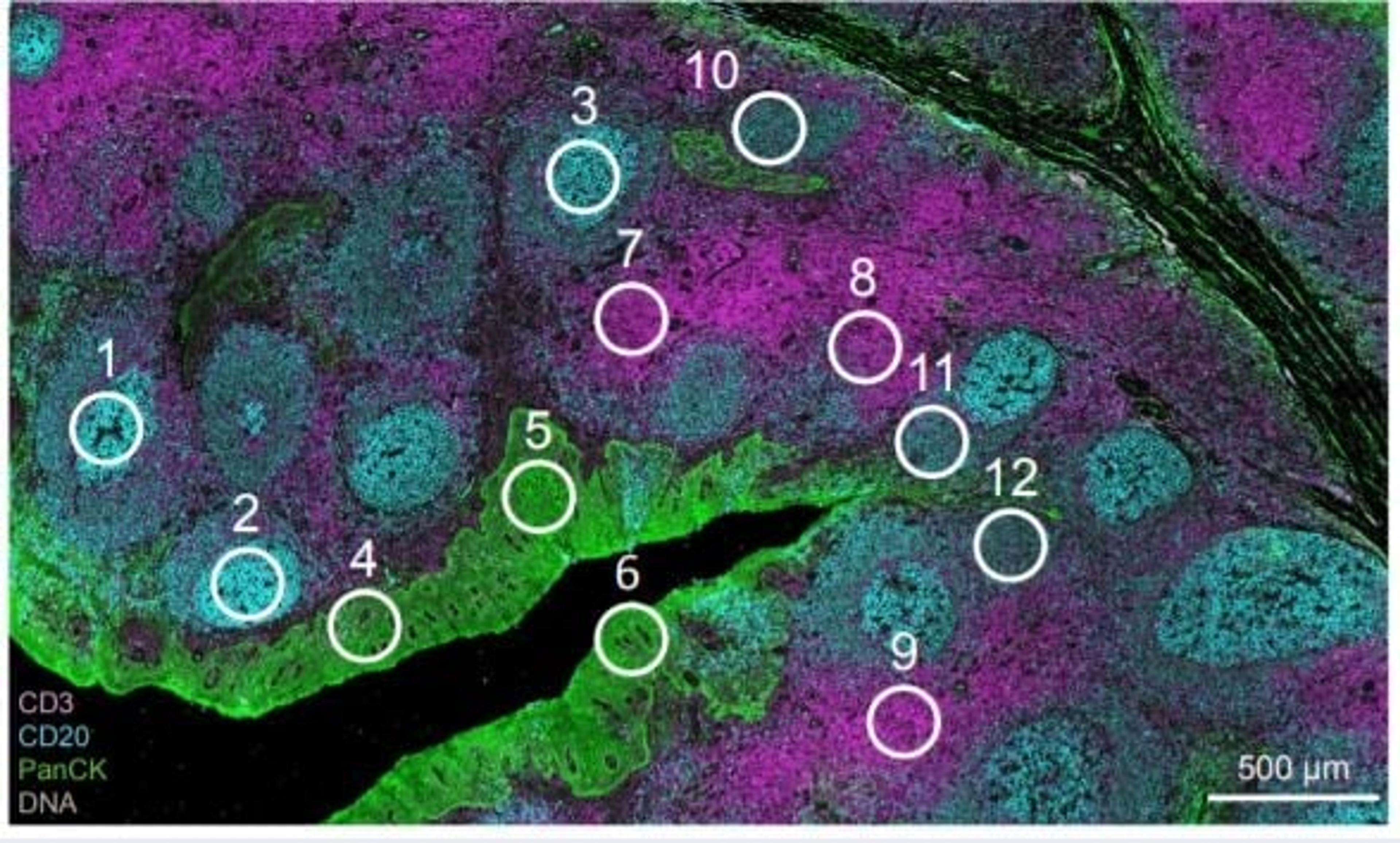
GeoMx Digital Spatial Profiler
Bruker Spatial BiologyMorphology driven high plex profiling on a single tissue section

CosMx™ SMI - Spatial Multiomics Single-Cell Imaging Platform
Bruker Spatial BiologyThe CosMx Spatial Molecular Imager is an FFPE-compatible, single-cell imaging solution that enables researchers to investigate intact tissues through the power of spatial multiomics.
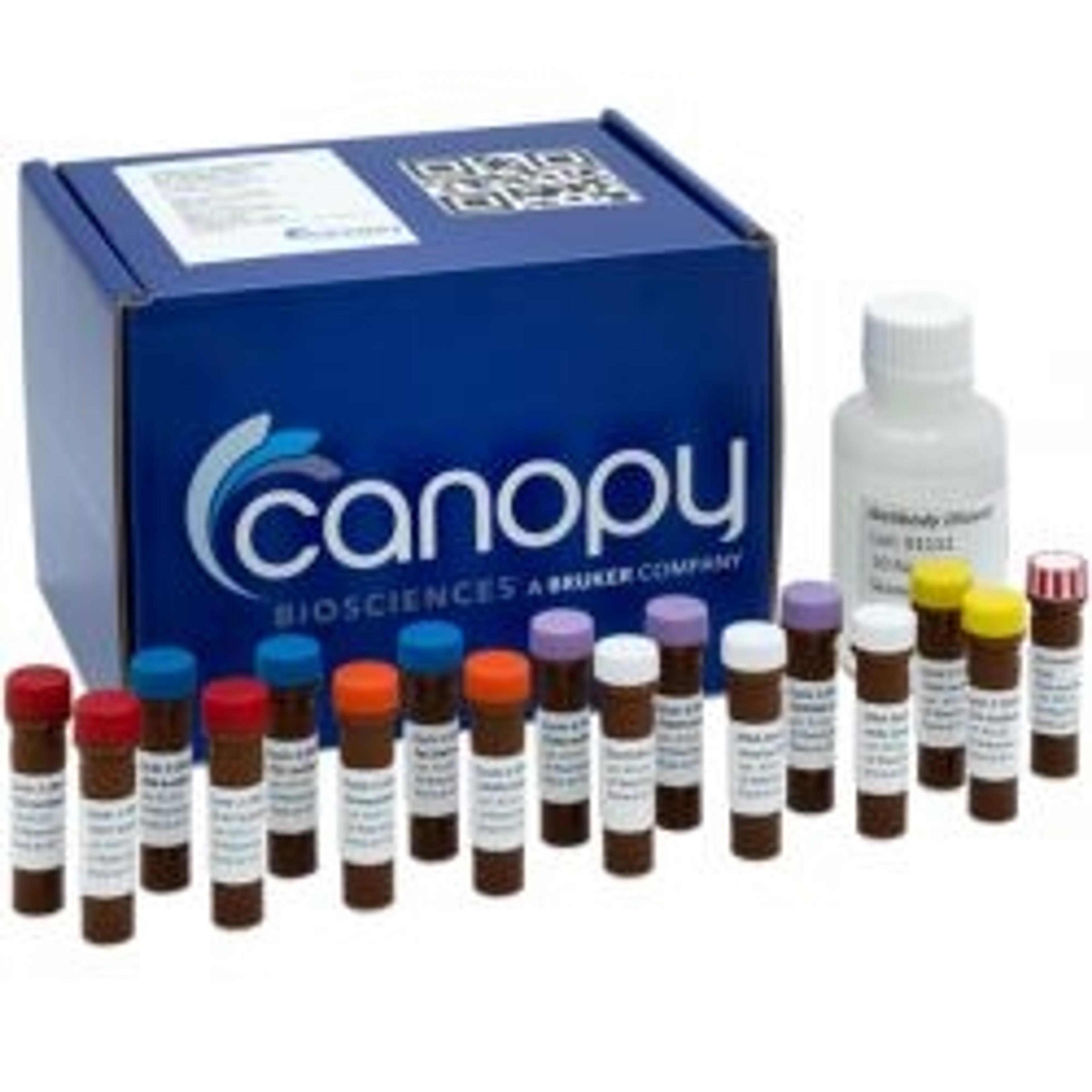
VistaPlex™ Assay Kits for ChipCytometry™
Bruker Spatial BiologyFast track precise spatial multiplexing