AQS3™delta
Advanced data processing software to leverage increased data quality
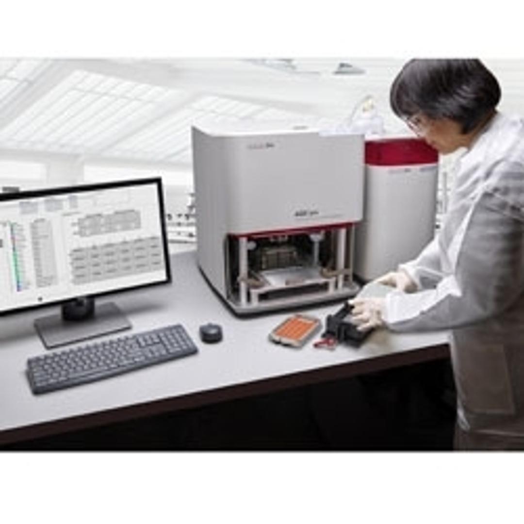
AQS3™delta software suite


The supplier does not provide quotations for this product through SelectScience. You can search for similar products in our Product Directory.
The AQS3delta software is a suite of analytical tools that within seconds turn high quality data into scientific insight.
Concerned about stability? Then track changes between spectra, at individual spectral locations or across structural motifs that you can define. Or analyze similarity with the Area of Overlap tool. Take a look at the data flow analysis below to see one way how the AQS3delta software processes and presents data to maximize the value of each measurement.

Analysis of an IgG1 Sample by Microfluidic Modulation Spectroscopy (MMS)
Microfluidic Modulation Spectrometry (MMS) is a novel protein characterization technique that combines a microfluidic cell and a tunable mid-IR quantum cascade laser source to assess the comparability, similarity, quantitation linearity, aggregation, and denaturation of proteins by analyzing the absorbance spectra and the high order structure (HOS). To evaluate the data quality and performance of MMS, an IgG1 sample was analyzed at different concentrations ranging from 0.1 mg/mL to 12.3 mg/mL using a RedShiftBio AQS3pro pre-production instrument.
Enhanced Protein Structural Characterization using Microfluidic Modulation Spectroscopy
RedShiftBio has developed a powerful new tool for protein structural analysis based on Microfluidic Modulation Spectroscopy (MMS). This technology shows significant increases in sensitivity, dynamic range, accuracy and utility for determination of protein secondary structure, quantitation, similarity, stability and aggregation. The analyzer utilizes a tunable mid-infrared quantum cascade laser to generate an optical signal 1000X brighter than conventional sources used in FTIR spectroscopy.
Early Events in Amyloid Formation by Lysozyme Detected by Microfluidic Modulation Spectroscopy
The self-assembly of lysozyme to form amyloid fibrils is associated with systemic amyloidosis, a disease characterized by the presence of amyloid deposits in various organs of the body. The early events associated in the self-assembly of lysozyme are not well understood. In this work, we used Microfluidic Modulation Spectroscopy (MMS) to characterize the early events in the self-assembly of human lysozyme. Through MMS, we were able to probe the mid-IR absorption band of the protein which is sensitive to both α-helix and β-structure. With heating at 60 ˚C, the β-turn content of the protein increases while its α-helical content decreases. This result suggests that the first structural transition in the self-assembly of human lysozyme is an α-helix to β-structure conformational rearrangement.