Establishing Phantoms to Improve MRI Signal and Quality

SelectScience® spoke to Dr Michael Boss, NIST, about his work to improve brain imaging

19 Feb 2016
Lois Manton-O'Byrne, PhD
Executive Editor

Editorial article

SelectScience® spoke to Dr Michael Boss, NIST, about his work to improve brain imaging

National Institute of Standards and Technology (NIST) NIST is a measurement standards laboratory, promoting U.S. innovation by advancing science, standards, and technology.

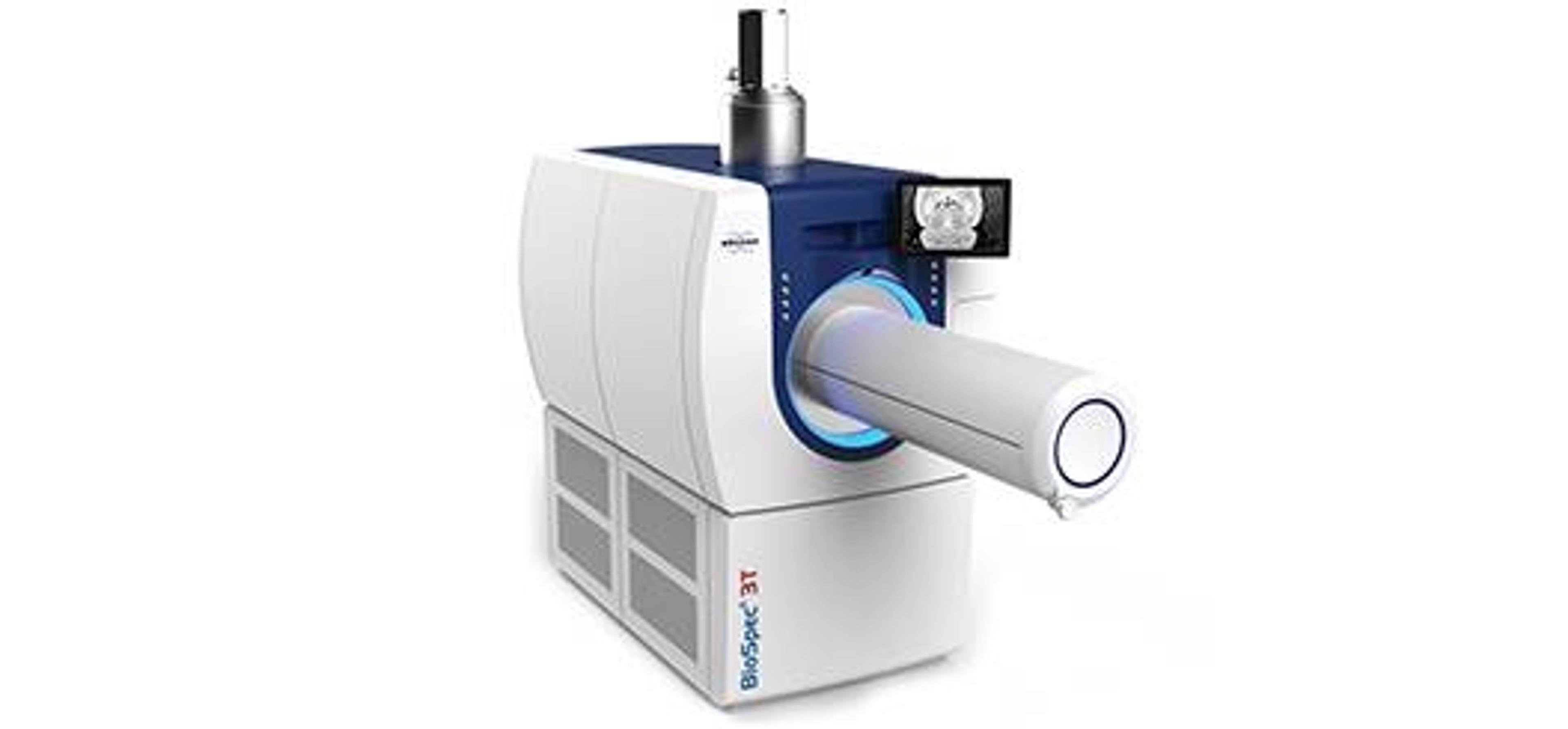
Nuclear magnetic resonance (NMR), is a physical phenomenon that can reveal detailed information on the chemical structure of unknown compounds, accurately measure purity, and, most commonly, be utilized in magnetic resonance imaging (MRI) for medical diagnosis. However, MRI information can be distorted or littered with artifacts, which lessen the certainty of a diagnosis. SelectScience spoke to Dr Michael Boss, of the Magnetic Imaging Group at NIST and specializing in brain imaging, who is working on reducing these interferences.

“Brain imaging provides a challenge”, Dr Boss explains; “neural tissue is very complex, as is the geometry of the head”. He is working towards the development of quantitative reference standards for MRI, and the establishment of a ground truth. “Ground truth is the true value of a physical parameter”, and without it, there is “no guarantee that the number measured is the true value”, he explained. Using the “gold standard” techniques of NMR, the accurate measurement of the ground truth will allow accurate measurements of all kind of parameters.

Eliminating distortions with phantoms

The standards Dr Boss is developing are known as “phantoms”. They are reference objects with known properties and can establish the “geometric distortion of an MR image, the signal-to-noise or contrast-to-noise ratio, or the spatial resolution”. The goal of his group is to “establish quality control protocols that enable reproducible, quantitative MRI. For example, if you were looking to generate a spatial map of the diffusion coefficient in a cancerous tumor, having a phantom with known diffusion properties is paramount to obtaining an accurate map in vivo.”

Magnetic resonance provides such an abundance of information that there exist almost limitless possibilities for harmonization with other spectroscopic and imaging techniques. Combining NMR/MRI with optical spectroscopic techniques could provide “a wealth of information on living subjects – using complementary but physically different techniques” allows for confident cross-validation. NIST is interested in combining MRI with positron emission tomography (PET) as a standard for multi-modality imaging. They are also planning on using microCT (another imaging technique utilizing x-rays) as an independent means of validating some of their phantom structures.

The future of MRI

“There are currently strongly efforts to push MRI to strengths of 7 tesla and beyond”, Dr Boss revealed. At present, MRI is clinically performed at 1.5 and 3 tesla, and although going to higher field strengths increases the signal, “it also introduces new problems in terms of image quality”. However, if these problems can be overcome, “these new MRI scanners could have higher spatial resolution, provide new means of generating contrast in an MR image, enable better MR spectroscopy, and lead to improved patient care through better diagnostics.”

Learn more about NMR and spectroscopy on our dedicated page, or read about how NMR can be utilized in other applications.

Links

Tags

NMR and EPR SpectroscopyNuclear magnetic resonance (NMR) spectroscopy is used to resolve the local chemical environment of atomic nuclei with spin, revealing information on molecular structure, dynamic processes and chemical reactions of organic molecules, from proteins to synthetics. Electron paramagnetic resonance (EPR) also known as electron spin resonance (ESR) spectroscopy is used to detect and quantify paramagnetic species in a sample, including free radicals as transition metal ions. By immersing the sample in a strong magnetic field, both NMR and EPR spectrometers probe the sample with either radio waves or microwaves respectively. A range of benchtop, solid-state and time domain NMR spectrometers & EPR spectrometers are available, as well as NMR tubes, NMR solvents, software, coils, and magnets. Find the best NMR & EPR equipment in our peer-reviewed product directory: compare products, check customer reviews and receive pricing direct from manufacturers.Cellular PathologyCellular Pathology deals with the microscopic analysis of tissue samples and cells. Sample preparation and processing includes fixation, staining, sectioning and slide mounting, using equipment such microtomes and cryostats. In choosing immunohistochemistry and immunocytochemistry kits, consider chromogens, staining method, antibodies, microscopes and imaging.X-Ray Diffraction and SpectroscopyX-Ray diffraction & spectroscopy are used in material characterization to discern the structure and elemental composition of a sample. X-Ray diffractometers (XRD) are superior instruments in elucidating the dimensional atomic structure of crystalline materials, including powders, thin films and single crystals. For large unit cells or ordered macromolecules, consider small-angle X-ray scattering (SAXS). X-ray spectroscopic techniques include X-ray fluorescence (XRF) and X-ray photoelectron spectroscopy (XPS), both providing simple and accurate methods for determining the elemental composition of a material. Energy dispersive (EDXRF) and wavelength dispersive (WDXRF) XRF spectrometers are available, as well as handheld/portable devices. High-resolution, 3D microstructure characterization of materials can be achieved with X-ray microscopes combining sub-micron resolution imaging with 3D computed tomography. Find the best XRD and XRF spectrometers in our peer-reviewed product directory: compare products, check customer reviews and receive pricing direct from manufacturers.Cancer DiagnosticsThere are a wide variety of diagnostic tests for cancer available, and this range continues to expand as our knowledge of cancer improves. Current diagnostic methods include biopsy, imaging and blood tests for known biomarkers. New methods in research development include liquid biopsies and cancer breathalyzers.NeurobiologyNeurobiology studies the biology of the nervous system, including the structure and function of neurons, glial cells, and their interactions. It is a key field in understanding neurological disorders such as Alzheimer�s, Parkinson�s, and schizophrenia. Research in neurobiology is essential for developing therapies for these conditions. Browse our peer-reviewed product directory to find the best neurobiology research tools, compare products, check reviews, and get pricing directly from manufacturers.Electron Magnetic ResonanceMRIPETDigital ImagingDigital imaging is a technique that captures images of biological and chemical samples in high resolution, often using advanced cameras and sensors. This technology is crucial in microscopy, diagnostics, and quality control. Explore digital imaging systems in our peer-reviewed product directory; compare products, check reviews, and get pricing directly from manufacturers.