ResourceLife Sciences
Understanding Immuno-Oncology with Real-Time Live-Cell Analysis
1 May 2019Immuno-oncology research requires the measurement of both tumor and immune cell activity, in isolation and together. This poster illustrates how these processes, from proliferation to phagocytosis, can be visualized in real time with the Incucyte® Live-Cell Analysis System from Sartorius.
Related products
Request Quote for All Products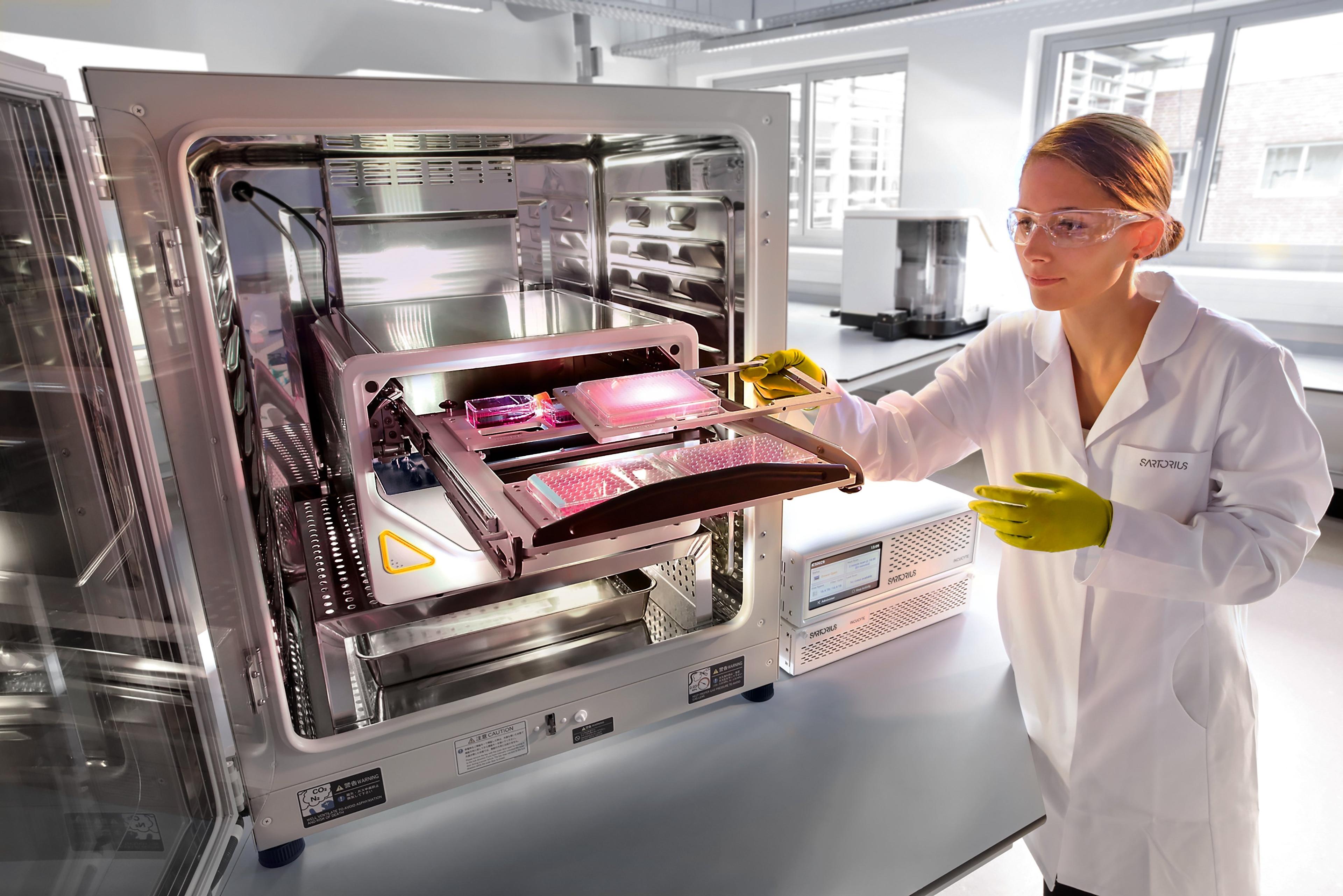
Incucyte® Live-Cell Analysis Systems
Sartorius GroupIncucyte ® , Empower Live-Cell Analysis Inside Your Incubator
Links
Tags
Cell / Tissue CultureCell culture or tissue culture is used to study the biology of cells or tissues and to isolate cellular products in an environment which can be manipulated and well defined. Accurately control your culture environment with bioreactors or culture incubators, bind your cells to a surface or together with an extracellular matrix. Distinguish cell types with differential media or proliferate cells with certain characteristics using selective media. Enrich your media with supplements such as growth factors, sera and vitamins. Find the best cell and tissue culture products, kits and equipment in our peer-reviewed product directory: compare products, check customer reviews and receive pricing direct from manufacturers.ImmunologyImmunological techniques measure and characterize immune responses. Immunology kits and analysis systems often use techniques such as ELISA, radioimmunoassay (RIA) and immunodiffusion assays, Immunohistochemistry, and flow cytometry. Immunologists use equipment such as flow Cytometers, plate readers, plate washers and fluorescent microscopes.Cellular PathologyCellular Pathology deals with the microscopic analysis of tissue samples and cells. Sample preparation and processing includes fixation, staining, sectioning and slide mounting, using equipment such microtomes and cryostats. In choosing immunohistochemistry and immunocytochemistry kits, consider chromogens, staining method, antibodies, microscopes and imaging.In Vivo Imaging Systems<i>In vivo</i> imaging systems, including pre-clinical imaging systems and medical imaging systems are used to non-invasively visualize and capture images of live animals and plants. Monitor the natural processes or diseases of your subjects using small-animal pre-clinical imaging systems, including single photon positron emission tomography (SPECT), positron emission tomography (PET), computed tomography (micro-CT), magnetic resonance imaging (MRI), X-ray radiography, ultrasound, fluorescence and bioluminescence imagers. Multimodal systems and software solutions are also available for correlative analysis of organ, tissue, cell, or molecular-level processes. Find the best in vivo imaging products in our peer-reviewed product directory: compare products, check customer reviews and receive pricing direct from manufacturers.Cell AnalysisThe analysis of cells allows researchers to understand the factors which contribute to cell health and function. These influencing processes can then be predicted and altered, leading to the development of medication and disease treatments.Live Cell ImagingLive cell imaging is the study of living cells using microscopes and high-content imaging systems. This technique provides in-depth insight into fast and complex biological processes, by allowing dynamic imaging of living cells instead of acquiring an individual image at a single point in time.TumorsTumor research focuses on understanding abnormal cell growth that leads to cancer. Identifying biomarkers, studying tumor microenvironments, and developing targeted therapies are critical for advancing cancer treatment. Early detection and personalized treatment options are key to improving outcomes for patients. Browse our peer-reviewed product directory to explore tools for tumor research, diagnostics, and cancer therapies; compare products, read customer reviews, and get pricing directly from manufacturers.