ResourceSpectroscopy
Speed Up Your Correlative Workflow with Shuttle & Find for ZEN Imaging Software
1 May 2016This guide explains how Shuttle & Find allows you to link functional data from light microscopy with information about the ultrastructural context revealed by electron microscopy. Fully integrated into ZEN imaging software, it controls all required functions of both the light and electron microscopes, enabling you to achieve an easy and intuitive workflow from instrument to instrument.
Related products
Request Quote for All Products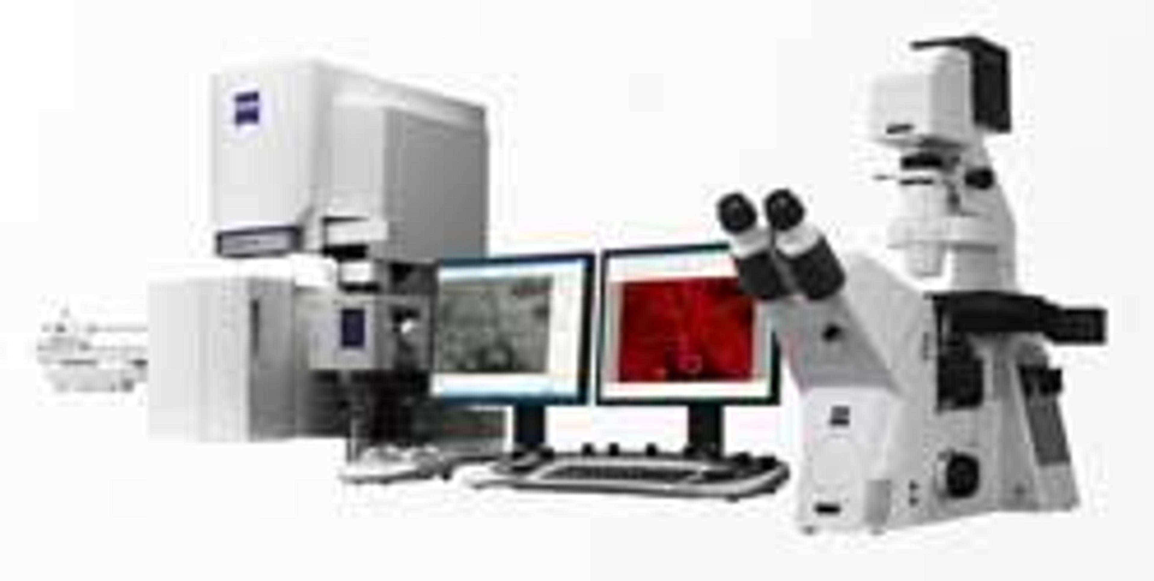
ZEISS Shuttle & Find for ZEN Imaging Software for Life Sciences
ZEISS Research Microscopy SolutionsSpeed Up Your Correlative Workflow.
Links
Tags
Sample PreparationSample preparation can improve the quality and speed of separation techniques. Products to assist sample preparation include filtration equipment, evaporators, membranes and sieves.Atomic Absorption / Emission SpectroscopyAtomic absorption spectroscopy (AAS) and atomic emission spectroscopy (AES) — also called optical emission spectroscopy (OES) — are used to detect the elemental constituents in samples. Both techniques involve the atomization of a sample. Atomic absorption spectrometers may use a flame or furnace to create an atomic vapor of the sample before irradiation with spectral light. Optical emission spectrometers may use a flame, inductively coupled plasma (ICP), microwave plasma (MP) or spark arcs to atomize and excite the sample. At higher excitation energies, electrons can be emitted instead of photons, which can be useful for samples that can’t be atomized and for surface analysis. Explore electron spectroscopy equipment such as Auger spectrometers and photoelectron spectrometers for surface elemental analysis of samples. Find the best atomic absorption, photoelectron and optical emission spectrometers in our peer-reviewed product directory: compare products, check customer reviews and receive pricing direct from manufacturers.Raman SpectroscopyRaman spectroscopy is used to discern the vibrational and rotational states of molecules and hence the chemical composition of a sample by measuring the inelastic scattering of monochromatic light. Explore a range of Raman spectrometers, including handheld/portable Raman spectrometers for QC/QA labs and in situ spectrometers for processes. Conduct Raman imaging for microanalysis of mixed samples using a Raman microscope. Raman spectrographs are also available. Find the best Raman spectroscopy products in our peer-reviewed product directory: compare products, check customer reviews and receive pricing direct from manufacturers.UV-Visible SpectroscopyUltraviolet-visible (UV-Vis) spectrophotometers are used to measure the interaction of UV and visible light with a sample, including transmission, reflectance & absorbance. The two major instrument classes are single-beam or double-beam spectrophotometers. More specialized equipment includes colorimeters, spectroradiometers and refractometers. Portable and microvolume spectrophotometers are also available. For the modular spectroscopy lab, explore a range of light sources for combination with a spectrograph/spectrometer and optics. Find the best UV-Vis spectroscopy products in our peer-reviewed product directory: compare products, check customer reviews and receive pricing direct from manufacturers.Gel Doc / Image AnalysisGel documentation (gel doc) or gel imaging systems are used for the analysis of proteins, antibodies and nucleic acid immobilized in polyacrylamide or agarose gels, membranes or microarrays. Explore a range of a gel imaging systems, densitometers, scanners, transilluminators or UV lamp + CCD cameras for your image analysis solutions. Colorimetric, fluorescent and/or radioisotopic samples can be visualized and documented for further analysis. See gel doc / Image analysis software for quantitative 1D and 2D analysis of your samples. Find the best gel doc / image analysis products in our peer-reviewed product directory: compare products, check customer reviews and receive pricing direct from manufacturers.Non-Destructive TechniquesNon-destructive techniques (NDT) describes a variety of analytical techniques used to evaluate the properties of a material. Common methods include ultrasonic, magnetic-particle, liquid penetrant, radiographic, remote visual inspection (RVI), and eddy-current testing. NDT is regularly used in forensic engineering, civil engineering, mechanical engineering, electrical engineering, systems engineering, aeronautical engineering, and medicine.Light MicroscopyLight microscopes or optical microscopes are used to visualize microscale objects under magnification, including cells, clinical specimens and materials. Lab equipment for light microscopy includes confocal microscopes, fluorescence microscopes, zoom and stereo microscopes. Microscope slides and imaging reagents are available for visualizing samples, as well as various microscope stages and incubators for large or temperature-sensitive samples. Find the best light microscopes in our peer-reviewed product directory: compare products, check customer reviews and receive pricing direct from manufacturers.Electron MicroscopyElectron microscopes (EM) are used to create high-resolution images of samples at the nanoscale by means of an accelerated beam of electrons as a source of illumination. Types of electron microscope include scanning electron microscopes (SEM), transmission electron microscopes (TEM), scanning transmission electron microscopes (STEM) and cryo-electron microscopes. Focused ion beam (FIB) microscopes are useful for modifying or milling a sample surface with nanometer precision, as well as imaging. Find the best electron microscopes in our peer-reviewed product directory: compare products, check customer reviews and receive pricing direct from manufacturers.Software PlatformsSoftware platforms are useful for various stages of laboratory experiments from data collection to data storage and processing. For instance lab software is available for system control, data management, data analysis and qualification / validation.Digital MicroscopyDigital microscopy involves using digital cameras and sensors to capture high-resolution images of samples for analysis. It offers enhanced imaging capabilities compared to traditional optical microscopy and is widely used in biological and material science research. Explore digital microscopy systems in our peer-reviewed product directory; compare products, check reviews, and get pricing directly from manufacturers.Cell ImagingCell imaging can be achieved using a number of techniques including confocal microscopy, transmission electron microscopy, atomic force microscopy, and light sheet microscopy.Cell AnalysisThe analysis of cells allows researchers to understand the factors which contribute to cell health and function. These influencing processes can then be predicted and altered, leading to the development of medication and disease treatments.ImagingImaging techniques are essential for obtaining visual representations of samples to understand structures, processes, and function in biological, chemical, and physical research. These tools range from traditional light microscopy to advanced imaging modalities like MRI and electron microscopy, providing researchers with valuable data for diagnostics, drug discovery, and material analysis. Explore imaging solutions in our peer-reviewed product directory to compare products, check reviews, and get pricing directly from manufacturers.MicroscopyMicroscopy is a technique used to observe small objects in detail, from cells to materials, using light or electron microscopes. It enables researchers to examine structures with high resolution, aiding in fields such as biology, medicine, and materials science. With advanced microscopy techniques, scientists can gain insights into cellular processes, tissue structures, and material properties. Explore the best microscopy solutions in our peer-reviewed product directory, compare products, read customer reviews, and get pricing directly from manufacturers.Correlative Microscopy