Set-Up of the CytoFLEX for Extracellular Vesicle Measurement
Set-Up of the CytoFLEX for Extracellular Vesicle Measurement
16 Aug 2015This application note describes how to set-up and standardize the CytoFLEX for particle measurement and discuss some pitfalls which should be avoided to get the best information from EVs detection. Extracellular vesicles are a heterogeneous cell-derived particle population in a size range between 50nm to 1,000nm. There is a growing interest not only from academic research groups to determine EV in several fluids such as cell culture supernatant, in plasma samples or in whole blood but also in clinical research since it has been shown that the measurement of microparticles (MPs) might be of clinical relevance. Compared with other methods of detection, flow cytometry has the big advantage that EV can be detected as rare events, in high numbers and by antigens on the surface, which characterize their cellular origin.
Related products
Request Quote for All Products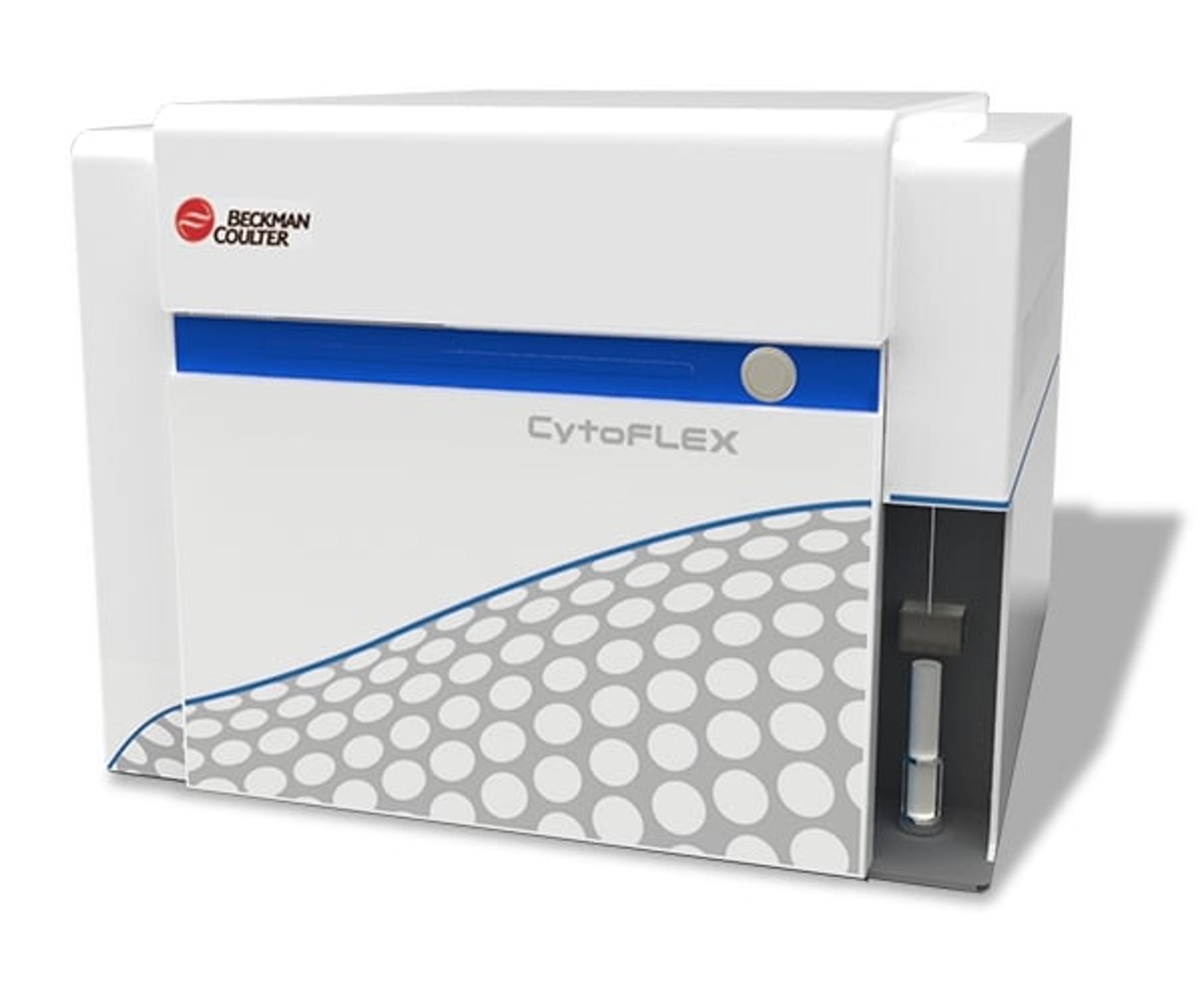
CytoFLEX Flow Cytometer
Beckman Coulter Life SciencesProviding quality and performance at any configuration, the CytoFLEX system provides powerful sensitivity and resolution for the simple to the most challenging applications. CytoFLEX delivers and surpasses capabilities expected in top tier analyzers, with excellent performance and nanoparticle resolution.