Raman Imaging of Monkey Brain Tissue
6 Mar 2014Fast and non-invasive methods for clinical and non clinical investigations for biological tissue are more and more required. This application note presents a method combining Raman spectroscopy and microscopy in a fully confocal instrument for the analysis of monkey brain tissue without chemical labeling.
Related products
Request Quote for All Products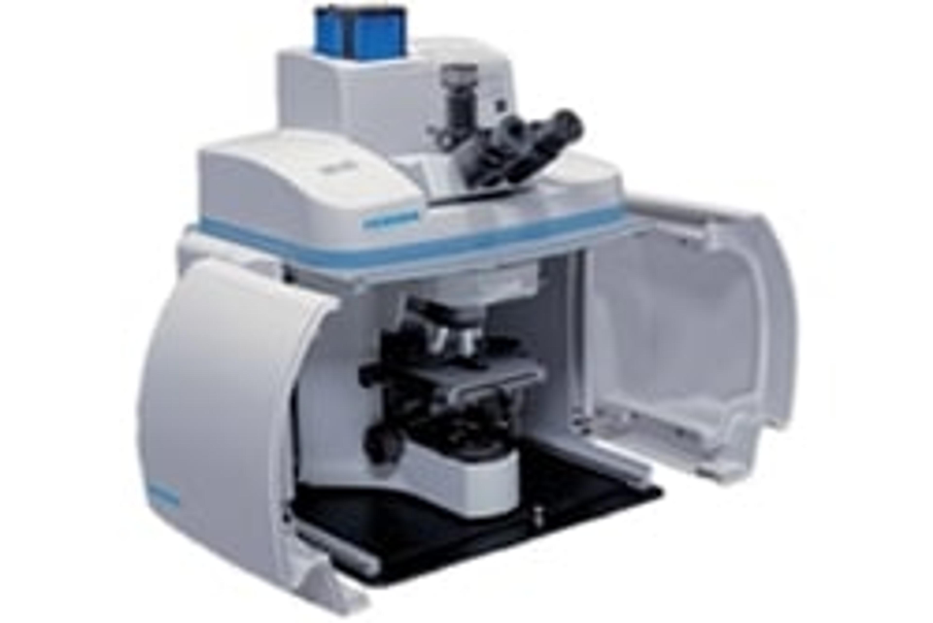
XploRA™ PLUS
HORIBA ScientificRaman imaging has never been so fast! Incorporating unique and powerful functions in a reliable, high performance system, ideally suited to the research and analytical lab, the XploRA PLUS is our best multi-sample, multi-user Raman microscope ever.It is fully confocal, not compromising image quality, spatial or depth resolution. The SWIFT Fast Raman images are the fastest fully confocal Raman images available, typically 10x faster than conventional Raman imaging.The simplicity and power of the XploRA PLUS is unmatched with an enhanced range of options such as multiple laser wavelengths, EMCCD detection, Raman polarisation and even Raman-AFM combination.XploRA™PLUS Features: SWIFTTM 10x faster Raman imaging Improved detection and sensitivity Full Confocality for complete image detail Full optical microscope so you can see your samples Maximum detail, resolution and range for enhanced spectroscopy HORIBA’s OneClick easy Raman analysis NIST traceable and patented Autocalibration options for validated results Ultimate optical stability- robust, reliable, long term operation Automated operation offering simple, powerful reliability 2 year base unit warranty as standard