ResourceLife Sciences
Python Blood Analysis by STEM
3 Dec 2018Understanding veterinary pathologies enables better conservation. This case study describes the analysis of python blood using cells prepared conventionally for electron microscopy, and thin sections subsequently imaged using an EVO15 HD SEM (scanning electron microscope) fitted with a transmission electron microscope (STEM) attachment.
Related products
Request Quote for All Products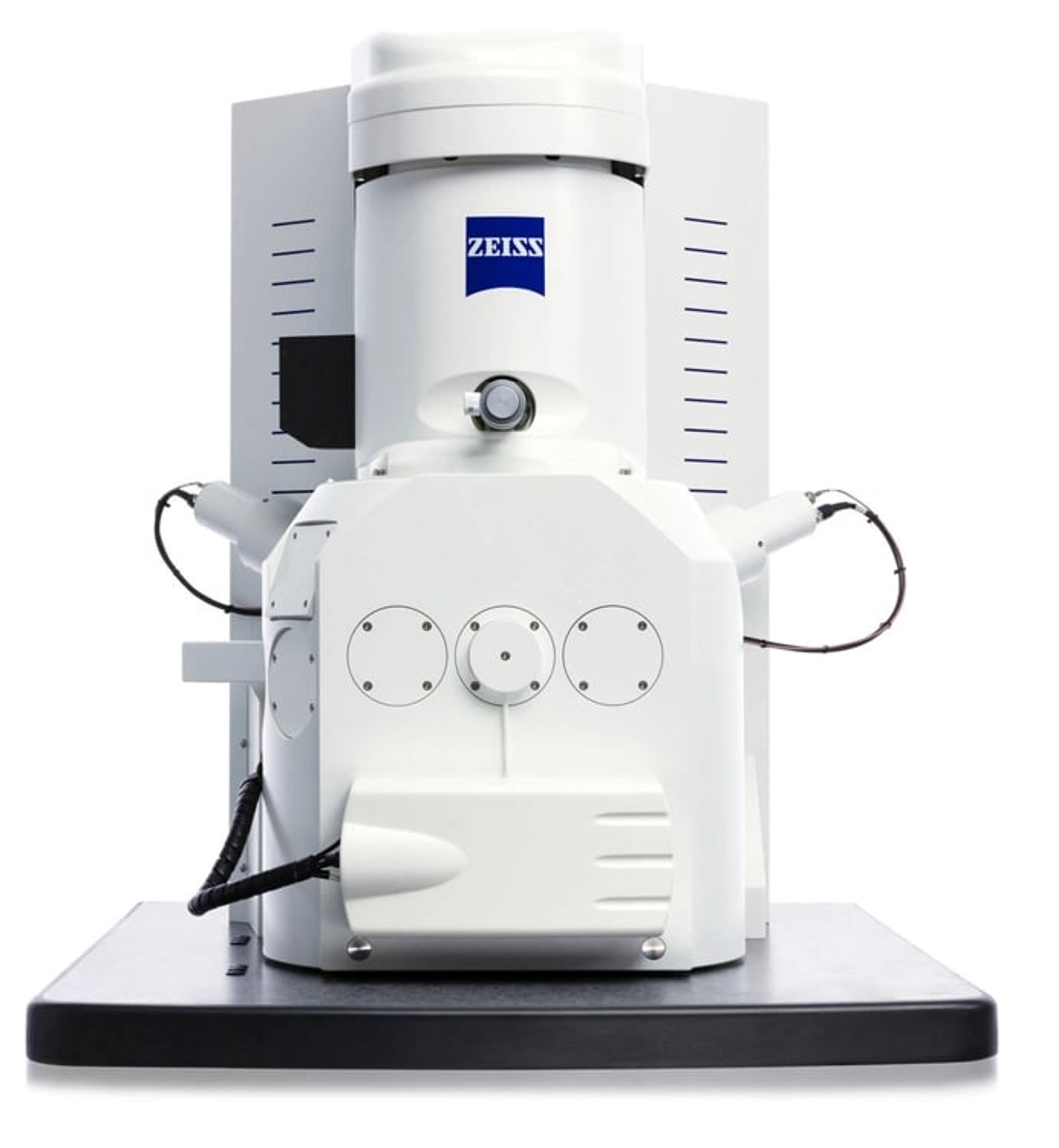
Links
Tags
Electron MicroscopyElectron microscopes (EM) are used to create high-resolution images of samples at the nanoscale by means of an accelerated beam of electrons as a source of illumination. Types of electron microscope include scanning electron microscopes (SEM), transmission electron microscopes (TEM), scanning transmission electron microscopes (STEM) and cryo-electron microscopes. Focused ion beam (FIB) microscopes are useful for modifying or milling a sample surface with nanometer precision, as well as imaging. Find the best electron microscopes in our peer-reviewed product directory: compare products, check customer reviews and receive pricing direct from manufacturers.Blood AnalysisThe analysis of blood is vital for many areas of life sciences and forensic investigations. Blood samples can be tested for a number of different reasons such as diagnosis, glucose levels, cholesterol and drug testing.