ResourceLife Sciences
Optical Sectioning Solutions for 3D Cancer Cell Analysis – Your Application Guide
30 Jun 2019In this eBook, we present several cancer-based examples of non-damaging, high-resolution, live-cell and fixed-cell assays, using some of the most innovative optical sectioning technologies available. These include the analysis of antibody-dependent cell-mediated toxicity (ADCC), 3D in-gel invasion, DNA damage signaling, mitochondrial uncoupling, and drug efficacy in multicellular models.
Related products
Request Quote for All Products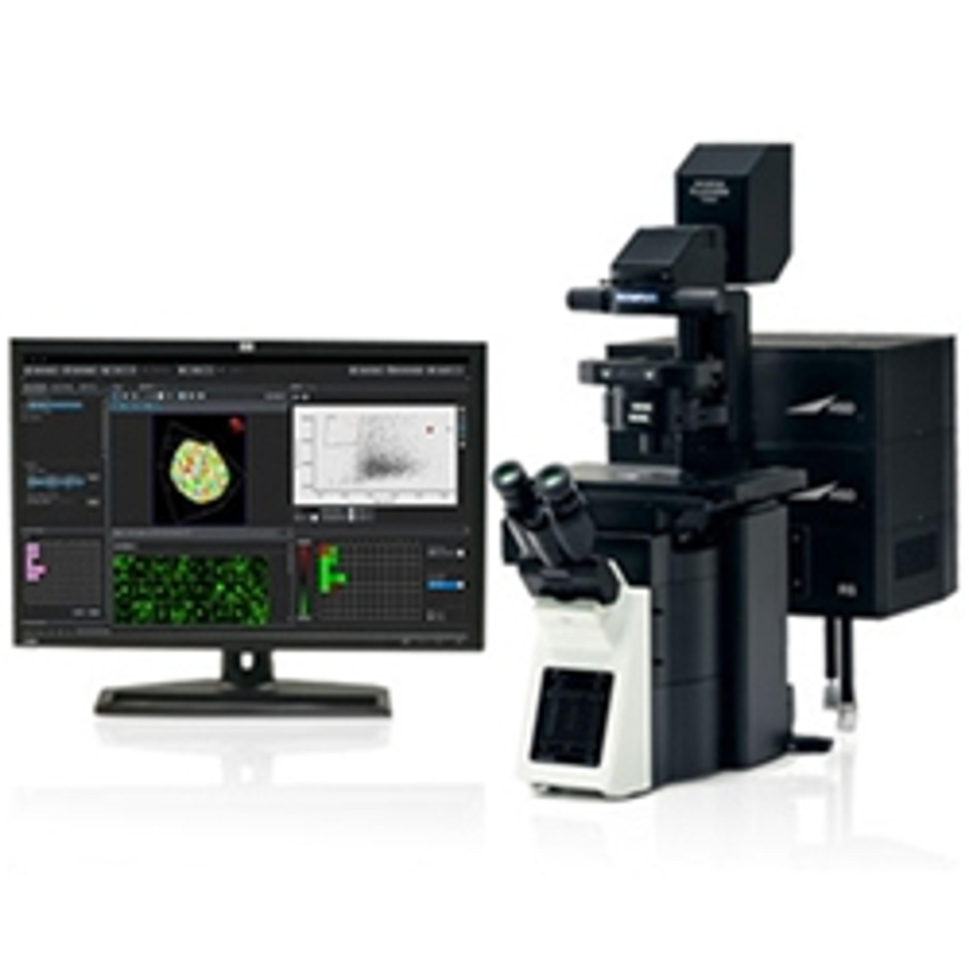
Links
Tags
Cell / Tissue CultureCell culture or tissue culture is used to study the biology of cells or tissues and to isolate cellular products in an environment which can be manipulated and well defined. Accurately control your culture environment with bioreactors or culture incubators, bind your cells to a surface or together with an extracellular matrix. Distinguish cell types with differential media or proliferate cells with certain characteristics using selective media. Enrich your media with supplements such as growth factors, sera and vitamins. Find the best cell and tissue culture products, kits and equipment in our peer-reviewed product directory: compare products, check customer reviews and receive pricing direct from manufacturers.Light MicroscopyLight microscopes or optical microscopes are used to visualize microscale objects under magnification, including cells, clinical specimens and materials. Lab equipment for light microscopy includes confocal microscopes, fluorescence microscopes, zoom and stereo microscopes. Microscope slides and imaging reagents are available for visualizing samples, as well as various microscope stages and incubators for large or temperature-sensitive samples. Find the best light microscopes in our peer-reviewed product directory: compare products, check customer reviews and receive pricing direct from manufacturers.3D Imaging3D imaging technologies allow for the visualization and analysis of three-dimensional structures at high resolution. These systems are used in fields like molecular biology, material science, and medical diagnostics. 3D imaging can be applied to visualize cells, tissues, and organs, providing valuable insights into their structure and function. Browse our peer-reviewed product directory to find the best 3D imaging solutions, compare products, check reviews, and get pricing directly from manufacturers.Live Cell ImagingLive cell imaging is the study of living cells using microscopes and high-content imaging systems. This technique provides in-depth insight into fast and complex biological processes, by allowing dynamic imaging of living cells instead of acquiring an individual image at a single point in time.MicroscopyMicroscopy is a technique used to observe small objects in detail, from cells to materials, using light or electron microscopes. It enables researchers to examine structures with high resolution, aiding in fields such as biology, medicine, and materials science. With advanced microscopy techniques, scientists can gain insights into cellular processes, tissue structures, and material properties. Explore the best microscopy solutions in our peer-reviewed product directory, compare products, read customer reviews, and get pricing directly from manufacturers.Cancer ResearchAlthough cancer is often referred to as a single condition, it actually consists of more than 100 different diseases. Microscopy, mass spectrometry, high throughput sequencing and flow cytometry are some of the most common techniques employed in cancer research labs.