ResourceLife Sciences
Observation of mitosis using Celloger Mini Plus
26 Sept 2022In this application note, the anti-mitotic activity of nocodazole against cancer cell line was examined by monitoring the cell division process. The cell division was monitored in real-time after treating the cell with or without nocodazole using the Celloger® Mini Plus.
Related products
Request Quote for All Products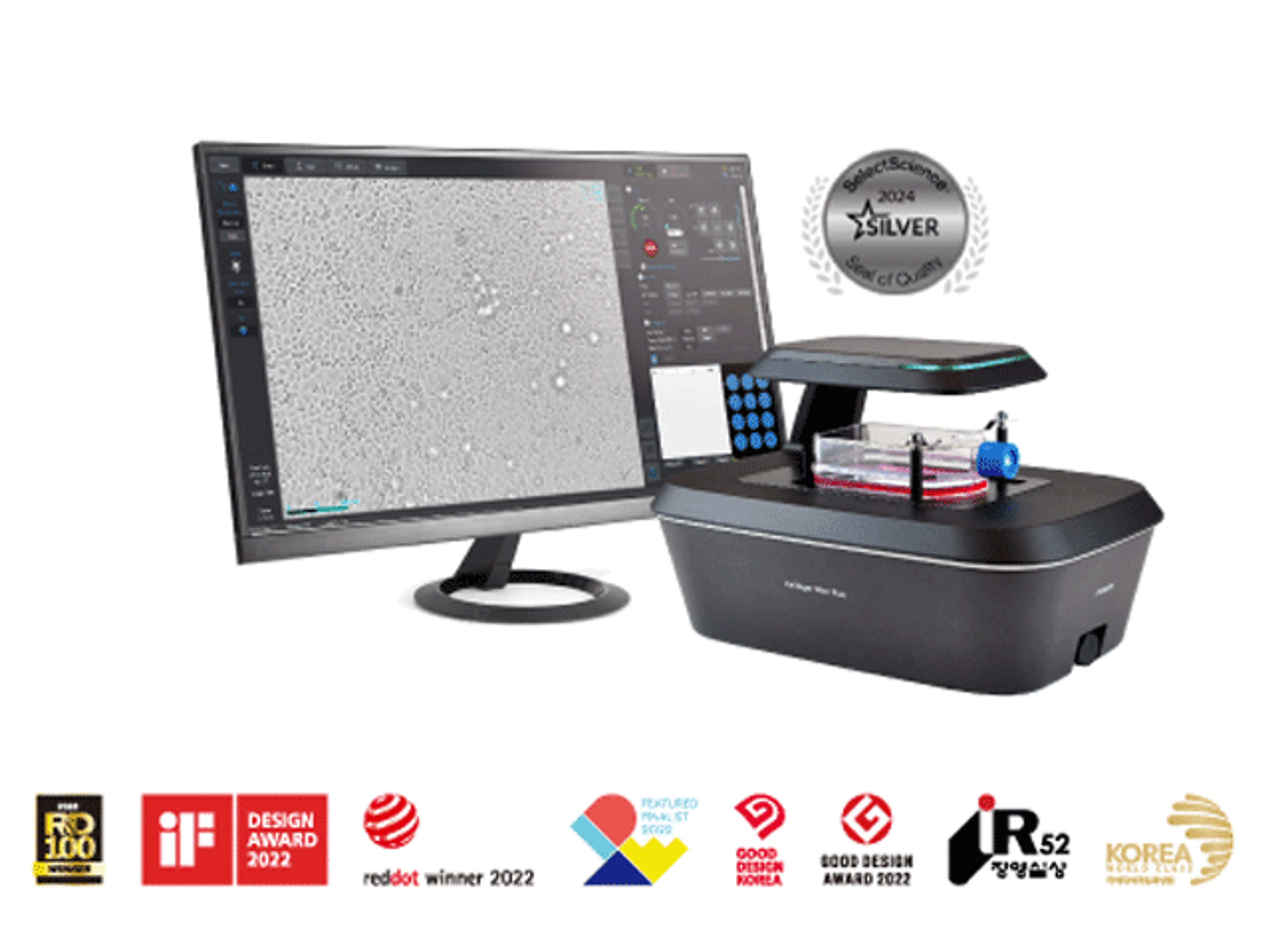
Celloger® Mini Plus, Automated Live Cell Imaging System from Curiosis
CURIOSISCelloger® Mini Plus is an automated live cell imaging system with fluorescence and brightfield microscopy. Celloger® Mini Plus makes it faster and easier to accumulate outstanding research results tailored to your research protocol.
Links
Tags
In Vivo Imaging Systems<i>In vivo</i> imaging systems, including pre-clinical imaging systems and medical imaging systems are used to non-invasively visualize and capture images of live animals and plants. Monitor the natural processes or diseases of your subjects using small-animal pre-clinical imaging systems, including single photon positron emission tomography (SPECT), positron emission tomography (PET), computed tomography (micro-CT), magnetic resonance imaging (MRI), X-ray radiography, ultrasound, fluorescence and bioluminescence imagers. Multimodal systems and software solutions are also available for correlative analysis of organ, tissue, cell, or molecular-level processes. Find the best in vivo imaging products in our peer-reviewed product directory: compare products, check customer reviews and receive pricing direct from manufacturers.Cancer CellsCancer cells are abnormal cells that divide uncontrollably, leading to the formation of tumors and the spread of cancer. Studying cancer cells is crucial for developing new treatments and understanding tumor biology. Explore cancer cell research products in our peer-reviewed product directory; compare products, check reviews, and get pricing directly from manufacturers.Live Cell ImagingLive cell imaging is the study of living cells using microscopes and high-content imaging systems. This technique provides in-depth insight into fast and complex biological processes, by allowing dynamic imaging of living cells instead of acquiring an individual image at a single point in time.