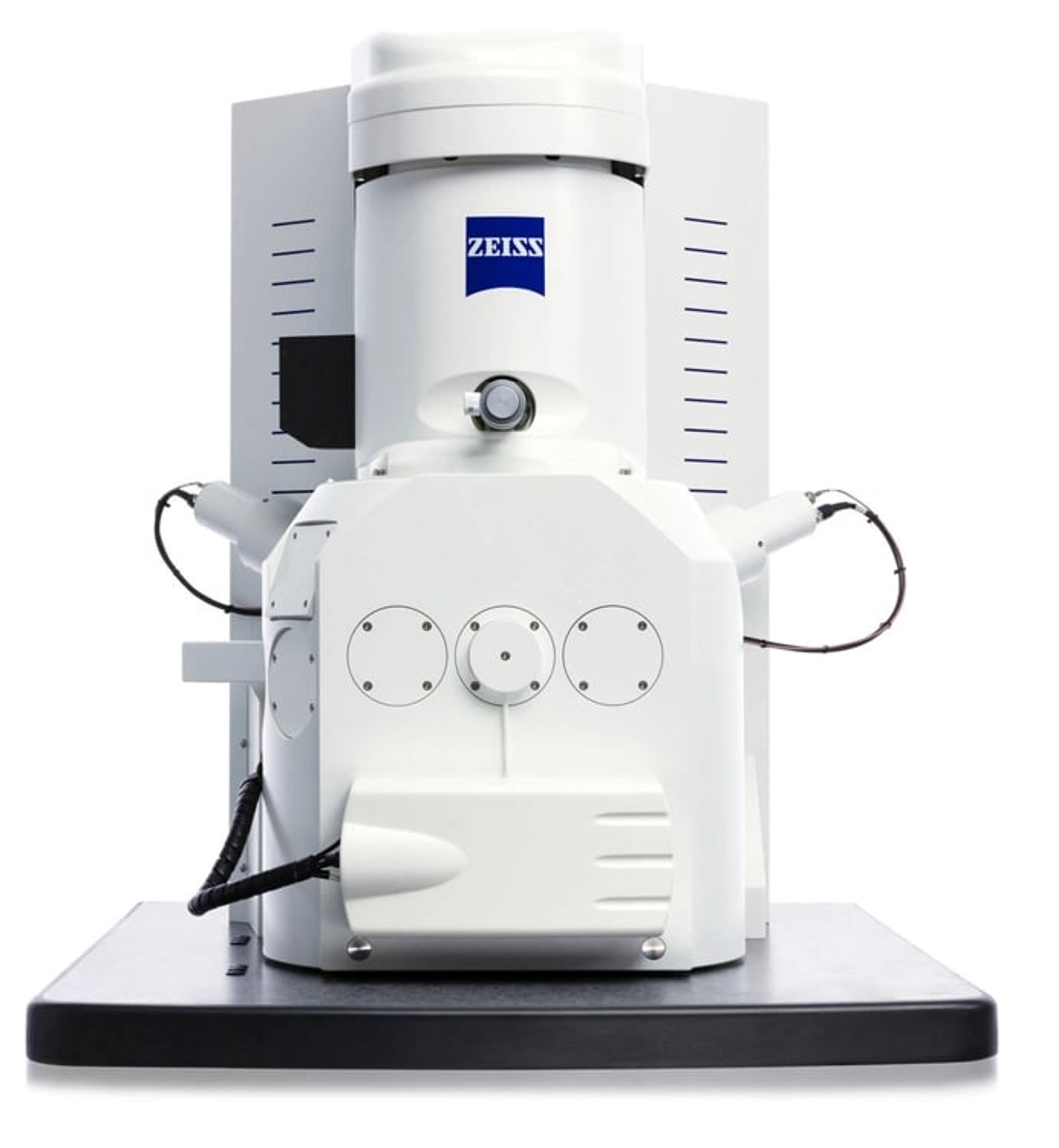
Multiscale Analysis of Bacteria Population in Legume Root Nodules with "Shuttle & Find"

4 May 2018

Correlative light and electron microscopy (CLEM) combines the overview information of fluorescence light microscopy (FLM) with structural details in scanning electron microscope (SEM) for high-content imaging. Hence, CLEM is very useful for investigating infection and colonization of the legume hosts by their rhizobial symbionts. This white paper illustrates a methodology involving the shuttle & find interface of CLEM to enable fast and reliable analysis of the bacterial infection of legume root nodule cells.

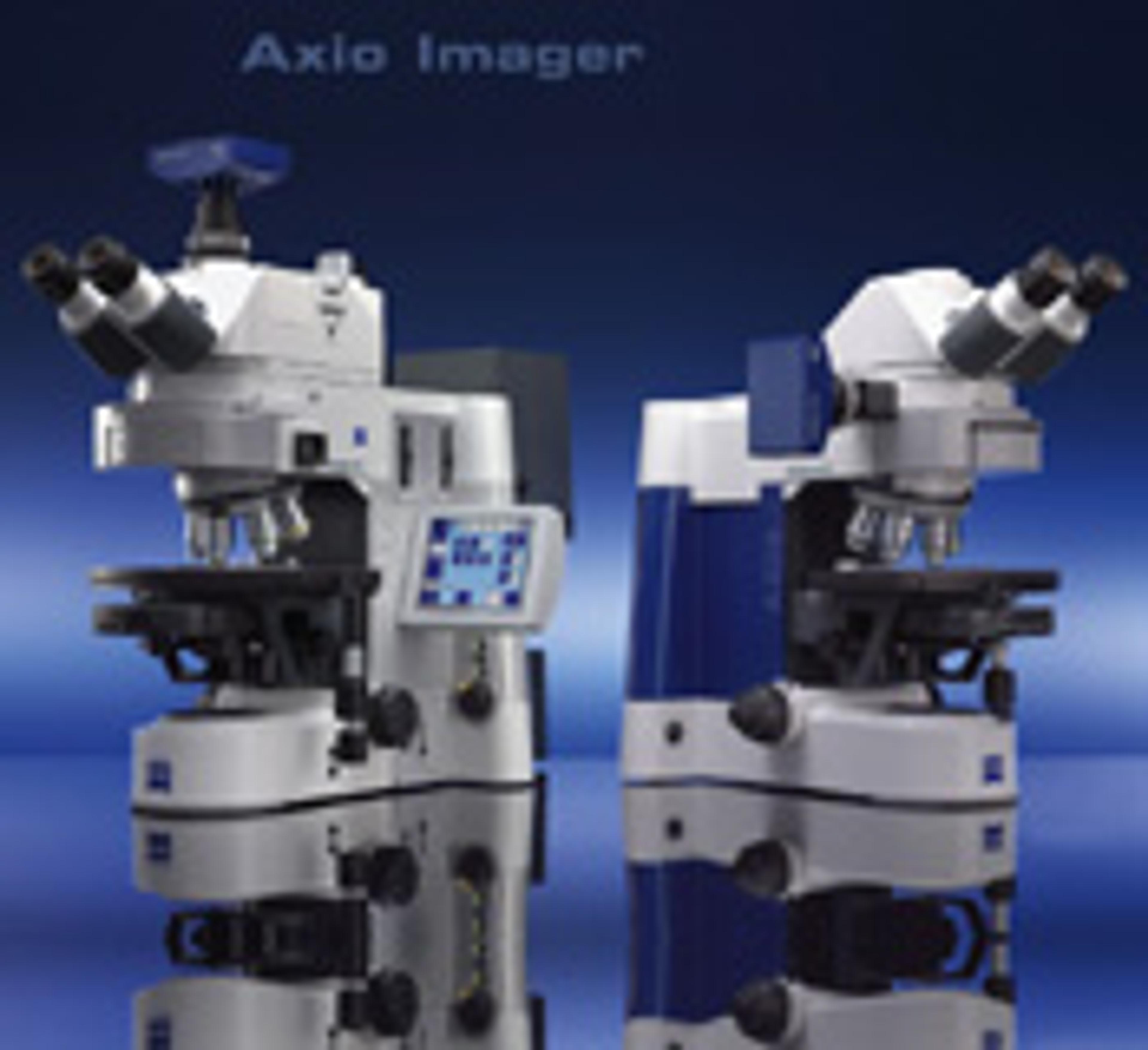
ZEISS Axio Imager - Modular System for Digital Fluorescence Microscopy

ZEISS Research Microscopy Solutions

Axio ImagerAn innovative modular system for digital fluorescence microscopy, featuring advanced flexibility and application versatility.  Featuring: New IC2S objectives (Infinity Contrast & Colour Corrected System) - optimise image quality and maximise contrast Special fluorescence filters - reduce exposure and image acquisition time for superior 3D imaging. 'Intelligent Stand' - automatically recognises added components.  "Contrast manager" ensures simple changes between contrasting techniques.  Integration: standard interfaces permit communication via USB and TCP/IP Vibration-free Imaging Cell - isolated within the stand for stable observation and unparalleled precision.  Apochromatic fluorescence beam path - ensures optimum colour correction Active stray light elimination - significantly higher contrast Fast, motorised reflector turrets - hold either six or ten filter modules.  HBO lamp: convenient, self-aligning homogeneous illumination LCI Plan-Neofluar objectives - live cell imaging Flexibility -  two freely configurable stand variants: Z1 (motorized) and D1(manual) or preconfigured stands M1 (motorized) and A1 (manual).  Rapid image acquisition with outstanding quality in up to 6 dimensions means more signal in less time.

(9)

Links

Tags

MicrobiologyMicrobiology is the study of microorganisms including protists, prokaryotes, fungi, and, often, viruses. Microorganisms are a useful research tool as genetic vectors and, in immunology, for antibiotic susceptibility testing, cellular biology and genetics. Microorganisms commonly grow readily in incubators with microbial culture media; this can contain chromogenic supplements to differentiate between cell lines. Estimate your culture’s density of microorganisms with colony counters, or screen and select colonies for desirable clones with automated colony pickers. Additionally, equipment is available to monitor environments for the presence of microbes and identify with microbial identification instruments. Find the best microbiology products in our peer-reviewed product directory: compare products, check customer reviews and receive pricing direct from manufacturers.Light MicroscopyLight microscopes or optical microscopes are used to visualize microscale objects under magnification, including cells, clinical specimens and materials. Lab equipment for light microscopy includes confocal microscopes, fluorescence microscopes, zoom and stereo microscopes. Microscope slides and imaging reagents are available for visualizing samples, as well as various microscope stages and incubators for large or temperature-sensitive samples. Find the best light microscopes in our peer-reviewed product directory: compare products, check customer reviews and receive pricing direct from manufacturers.Electron MicroscopyElectron microscopes (EM) are used to create high-resolution images of samples at the nanoscale by means of an accelerated beam of electrons as a source of illumination. Types of electron microscope include scanning electron microscopes (SEM), transmission electron microscopes (TEM), scanning transmission electron microscopes (STEM) and cryo-electron microscopes. Focused ion beam (FIB) microscopes are useful for modifying or milling a sample surface with nanometer precision, as well as imaging. Find the best electron microscopes in our peer-reviewed product directory: compare products, check customer reviews and receive pricing direct from manufacturers.ImagingImaging techniques are essential for obtaining visual representations of samples to understand structures, processes, and function in biological, chemical, and physical research. These tools range from traditional light microscopy to advanced imaging modalities like MRI and electron microscopy, providing researchers with valuable data for diagnostics, drug discovery, and material analysis. Explore imaging solutions in our peer-reviewed product directory to compare products, check reviews, and get pricing directly from manufacturers.Plant SciencePlant science studies the biology of plants, including genetics, physiology, ecology, and biotechnology. Research in this field is essential for improving crop yields, disease resistance, and sustainability. Explore plant science tools in our peer-reviewed product directory; compare products, check reviews, and get pricing directly from manufacturers.High Content ImagingHigh content imaging is a method combining two or more fluorescent microscopy experiments to identify substances that alter a cell’s phenotype in a desired manner. The process is adapted to multi-well plates and both the image acquisition and analysis are automated.MicrobiomeMicrobiome research focuses on the complex communities of microorganisms living in and on humans, animals, plants, and environments. It plays a crucial role in health, disease, and ecological balance. Explore tools and resources for microbiome analysis, including sequencing platforms and bioinformatics software.Bacterial DetectionBacterial detection uses methods such as cell culture, microscopy, and biochemical assays to identify harmful or beneficial bacteria based on their morphological and metabolic charactersistics. These techniques support disease diagnostics, food safety, and microbial research. Explore bacterial detection systems, kits, and consumables in our directory to compare products and read reviews.Correlative Microscopy