ResourceSpectroscopy

Multiple, Sequential DESI Images from a Single Tissue Section at Different Spatial Resolution

Multiple, Sequential DESI Images from a Single Tissue Section at Different Spatial Resolution

4 Feb 2016

This application from Waters Corporation demonstrate that desorption electrospray ionization (DESI) imaging can be utilized as a non-destructive imaging technique. This allows multiple analyses on the same tissue section at different spatial resolution.

SYNAPT G2-Si MS

Waters

Transform your lab's discovery capability with the SYNAPT G2-Si MS. Information. Informatics. Impact. SYNAPT enables extensive characterization of complex mixtures and molecules with uncompromising qualitative and quantitative performance, streamlined workflows and unparalleled platform versatility. With the SYNAPT G2-Si you get ultimate UPLC/MS/MS performance, powerful data independent & data dependant solutions, CID and ETD fragmentation capabilities, and a wide range of experimental options.

(5)

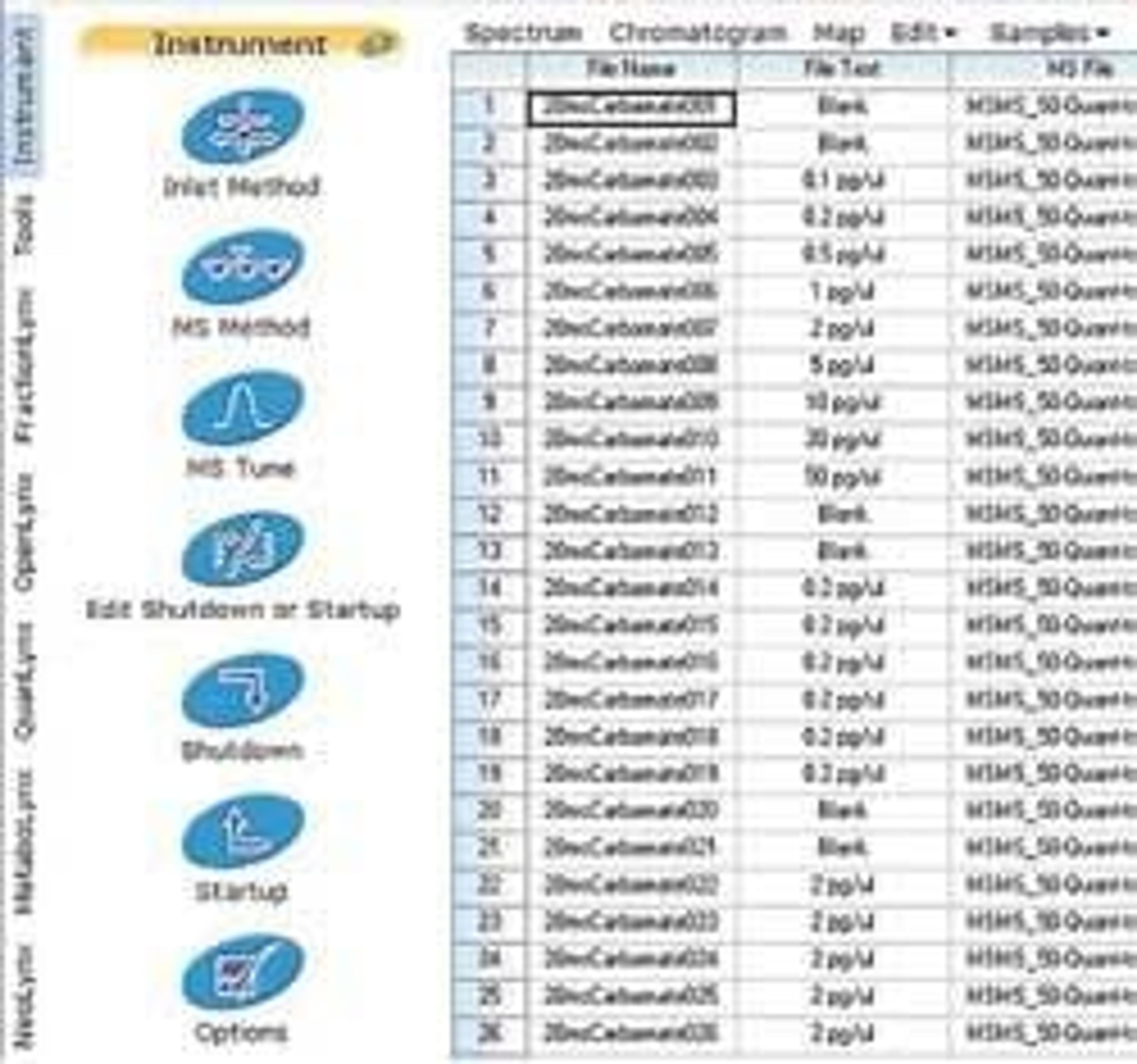
MassLynx MS Software

Waters

Acquire, Analyze, Manage, and Share Mass Spectrometry Information MassLynx™ Software increases the speed at which you can convert your sample data into valuable knowledge. It provides you with the fundamental platform to acquire, analyze, manage, and share your mass spectrometry information. MassLynx intelligently controls any Waters mass spectrometry system, from sample and solvent management components to mass spectrometer and auxiliary detectors. MassLynx Software may acquire nominal mass, exact mass, MS/MS and exact mass MS/MS data. MassLynx’s Sample List is the core of the system. It maintains and consolidates the data on all of your samples. You also initiate any activities related to your sample from the Sample List. This "sample centric" approach simplifies the interaction with your LC/MS or GC/MS system, acquired data and processed results. Process Application-specific Data MassLynx Software features general purpose and specialized Application Managers that provide information for your specific MS analyses and data. Two come standard with MassLynx: QuanLynx™ for automated quantification included as standard with MassLynx OpenLynx™ for qualitative screening and identification Optional Application Managers can perform: Targeted quantitative analysis Mass-directed purification Metabolite identification Deconvolution of complex chromatograms Metabonomics & metabolomics Protein identification and protein characterization

(8)

Links

Tags

Mass SpectrometryMass spectrometry (MS) is a powerful analytical technique used to identify and quantify molecules based on the mass-to-charge ratio of gas-phase ions. It provides detailed information about the structure, composition, and properties of compounds and is widely used across fields such as environmental monitoring, materials science, drug discovery and development, food and beverage testing, and wider chemical research. Key MS techniques include tandem mass spectrometry (MS/MS), liquid chromatography–mass spectrometry (LS-MS) and inductively coupled plasma (ICP-MS). Choosing from these wide range of techniques and technologies can be a daunting task, so keep up to date with scientific applications, performance expectations, and customer reviews here all in one place. Visit our product directory to receive quotes direct from the manufacturer. Gel Doc / Image AnalysisGel documentation (gel doc) or gel imaging systems are used for the analysis of proteins, antibodies and nucleic acid immobilized in polyacrylamide or agarose gels, membranes or microarrays. Explore a range of a gel imaging systems, densitometers, scanners, transilluminators or UV lamp + CCD cameras for your image analysis solutions. Colorimetric, fluorescent and/or radioisotopic samples can be visualized and documented for further analysis. See gel doc / Image analysis software for quantitative 1D and 2D analysis of your samples. Find the best gel doc / image analysis products in our peer-reviewed product directory: compare products, check customer reviews and receive pricing direct from manufacturers.Cellular PathologyCellular Pathology deals with the microscopic analysis of tissue samples and cells. Sample preparation and processing includes fixation, staining, sectioning and slide mounting, using equipment such microtomes and cryostats. In choosing immunohistochemistry and immunocytochemistry kits, consider chromogens, staining method, antibodies, microscopes and imaging.In Vivo Imaging Systems<i>In vivo</i> imaging systems, including pre-clinical imaging systems and medical imaging systems are used to non-invasively visualize and capture images of live animals and plants. Monitor the natural processes or diseases of your subjects using small-animal pre-clinical imaging systems, including single photon positron emission tomography (SPECT), positron emission tomography (PET), computed tomography (micro-CT), magnetic resonance imaging (MRI), X-ray radiography, ultrasound, fluorescence and bioluminescence imagers. Multimodal systems and software solutions are also available for correlative analysis of organ, tissue, cell, or molecular-level processes. Find the best in vivo imaging products in our peer-reviewed product directory: compare products, check customer reviews and receive pricing direct from manufacturers.Clinical MicrobiologyMicrobiology is the study of microorganisms including protists, prokaryotes, fungi, and, often, viruses. Microorganisms are a useful research tool as genetic vectors and, in immunology, for antibiotic susceptibility testing, cellular biology and genetics. Microorganisms commonly grow readily in incubators with microbial culture media; this can contain chromogenic supplements to differentiate between cell lines. Estimate your culture’s density of microorganisms with colony counters, or screen and select colonies for desirable clones with automated colony pickers. Additionally, equipment is available to monitor environments for the presence of microbes and identify with microbial identification instruments. Find the best microbiology products in our peer-reviewed product directory: compare products, check customer reviews and receive pricing direct from manufacturers.Tissue ProcessingTissue processing involves preparing biological tissue samples for microscopic analysis, including fixation, embedding, and sectioning. This process is essential for histopathology and diagnostic applications. Explore tissue processing products in our peer-reviewed product directory; compare products, check reviews, and get pricing directly from manufacturers.ImagingImaging techniques are essential for obtaining visual representations of samples to understand structures, processes, and function in biological, chemical, and physical research. These tools range from traditional light microscopy to advanced imaging modalities like MRI and electron microscopy, providing researchers with valuable data for diagnostics, drug discovery, and material analysis. Explore imaging solutions in our peer-reviewed product directory to compare products, check reviews, and get pricing directly from manufacturers.SpectrometrySpectrometry is a technique used to measure the mass-to-charge ratio of ions. It provides valuable information about the composition, structure, and molecular weight of compounds. Used across a variety of fields, including chemistry, biology, and environmental science, spectrometry is essential for identifying substances, analyzing mixtures, and developing new products. Explore the best spectrometry tools in our peer-reviewed product directory; compare products, check reviews, and get pricing directly from manufacturers.Multispectral Imaging