Modeling Cardiac Hypertrophy: Endothelin-1 Induction with High Content Analysis
11 Jul 2013This Application Protocol describes how to utilize iCell Cardiomyocytes in a 5-day post-plating in vitro cellular model of cardiac hypertrophy that is induced by endothelin-1 (ET-1) and quantified via increased BNP expression using a screening-compatible 384-well assay with high content analysis.
Related products
Request Quote for All Products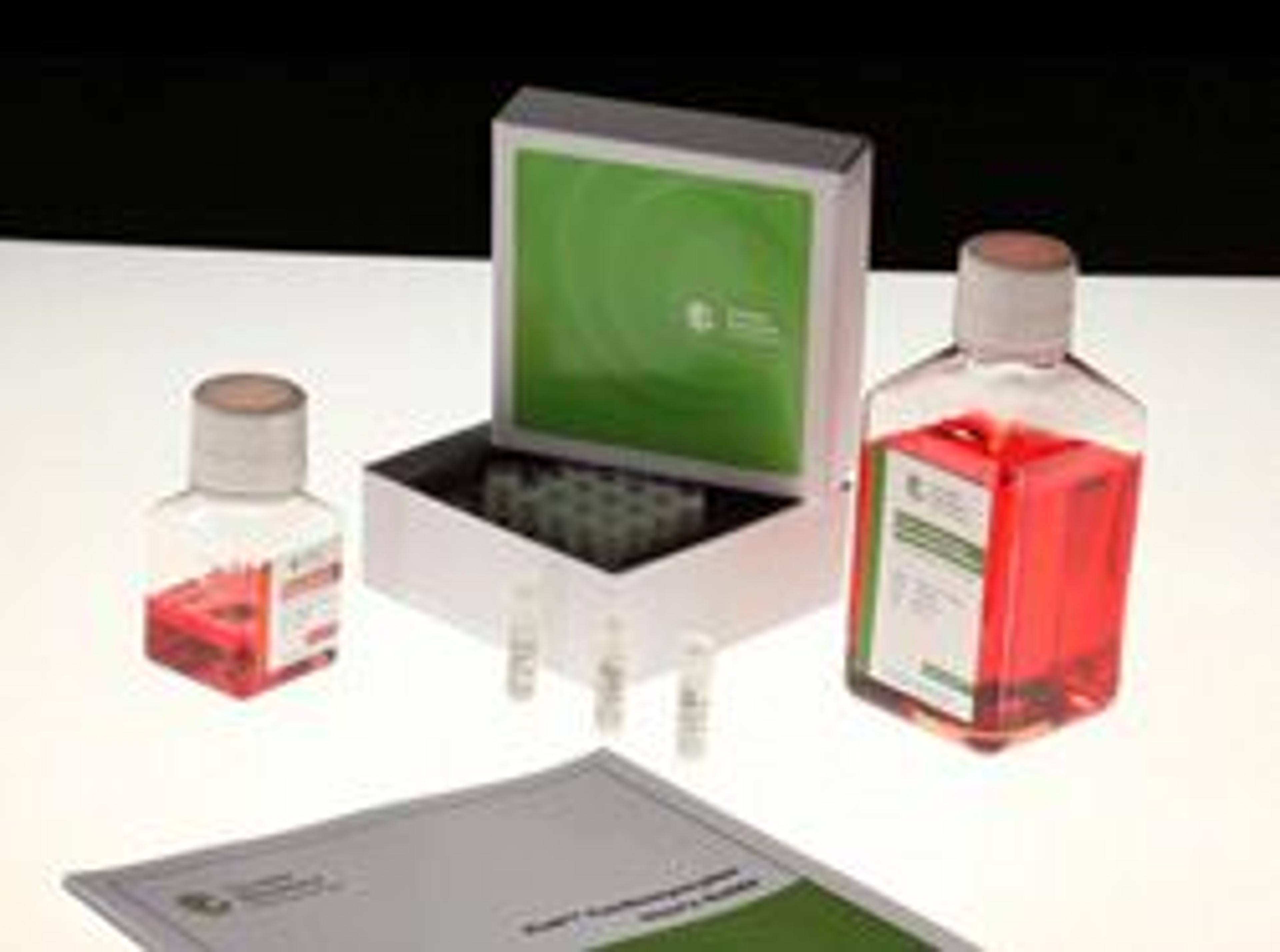
iCell Cardiomyocytes
Cellular Dynamics InternationaliCell® Cardiomyocytes, human induced pluripotent stem (iPS) cell-derived cardiomyocytes, aid drug discovery and improve the predictability of drug efficacy and toxicity screens, weeding out ineffective and potentially toxic compounds early in the pharmaceutical pipeline process before significant time and resources have been invested. iCell Cardiomyocytes are a mixture of spontaneously electrically active atrial, nodal, and ventricular-like myocytes that possess typical electrophysiological characteristics and exhibit expected electrophysiological and biochemical responses upon exposure to exogenous agents. Thus, these cells are a reliable source of human cardiomyocytes suitable for use in targeted drug discovery, toxicity testing, and other life science research. iCell Cardiomyocytes are shipped as cryopreserved suspensions of dissociated cells with specifically formulated culture media for optimal cell performance. Once thawed, iCell Cardiomyocytes remain viable in culture for up to two weeks, allowing for acute and chronic studies. iCell Cardiomyocytes Benefits: Human Cells - Saves valuable time, resources, and compound. Highly Pure Cell Population - Provides cardiac-specific response to reference molecules. Homogenous and Reproducible Fully Functional Model Acute and Longer-term Testing - Remain viable in culture for up to two weeks. iPS Cell-derived iCell Cardiomyocytes Applications: Cell-based Assays - Cell viability, Apoptosis, ATP production, Oxidative stress, Mitochondrial dysfunction. Electophysiological Applications - Conventional patch clamp recording, Microelectrode assay (MEA) recording.