Microscopy in the Field of Tool Engineering - the Microstructure of Hard Metal Drills
Microscopy in the Field of Tool Engineering - the Microstructure of Hard Metal Drills
10 Sept 2015The combination of microscopic and quantitative micro-structural analysis is a powerful method of obtaining information on geometry, usage properties and wear conditions of tools. The mechanical and usage properties of hard metal tools are crucially affected by their microstructure; therefore microscopic investigation of the micro- and macro-structure of the tools is a powerful and fast technique for checking the quality and corresponding usage properties of tools such as drills. This application notes presents possible applications of different microscopy systems from optical light to electron microscopy.
Related products
Request Quote for All Products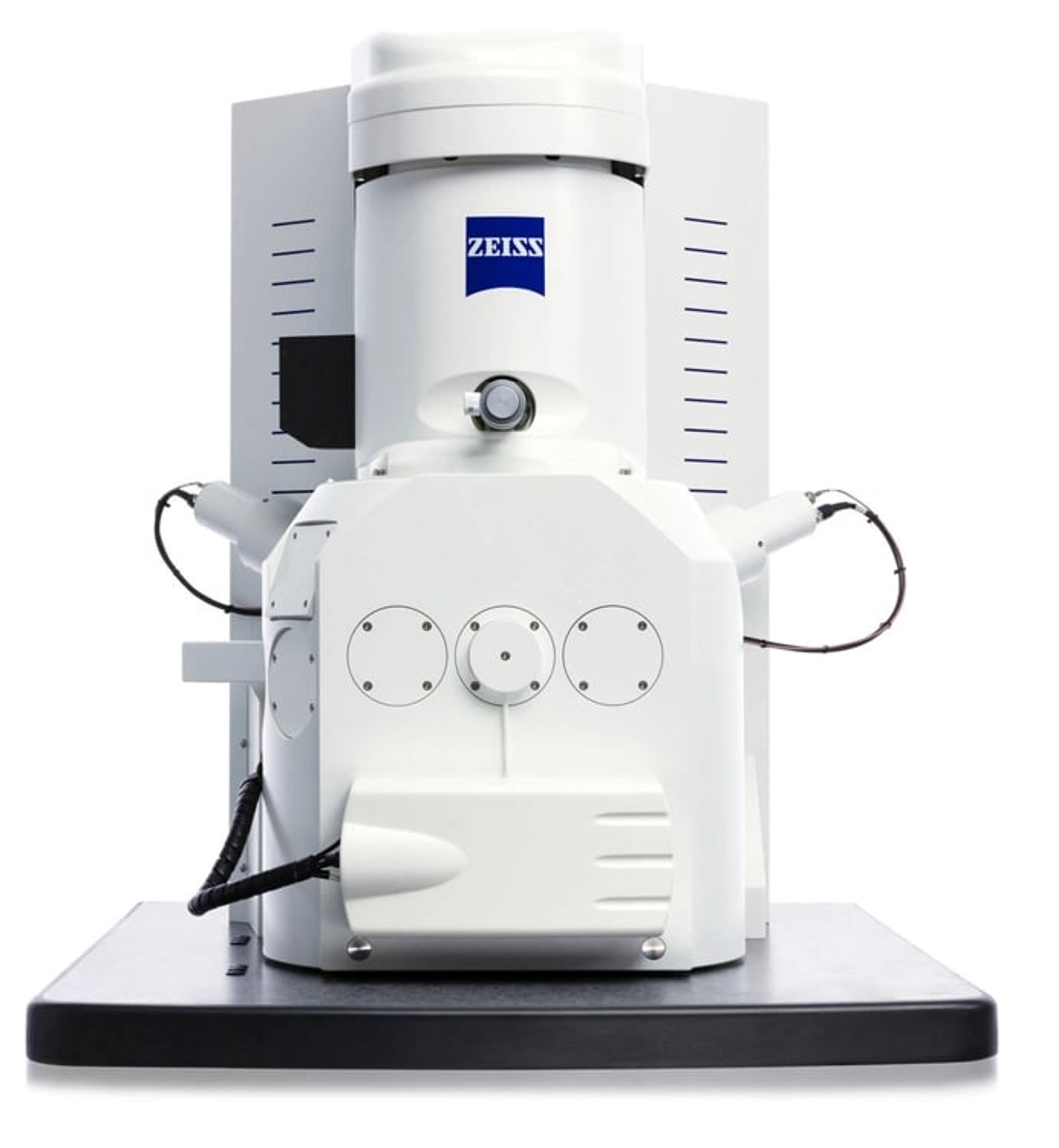

ZEISS Smartzoom 5
ZEISS Research Microscopy SolutionsSampling Made Simple: Your Automated Digital Microscope for Routine And Failure Analyses.
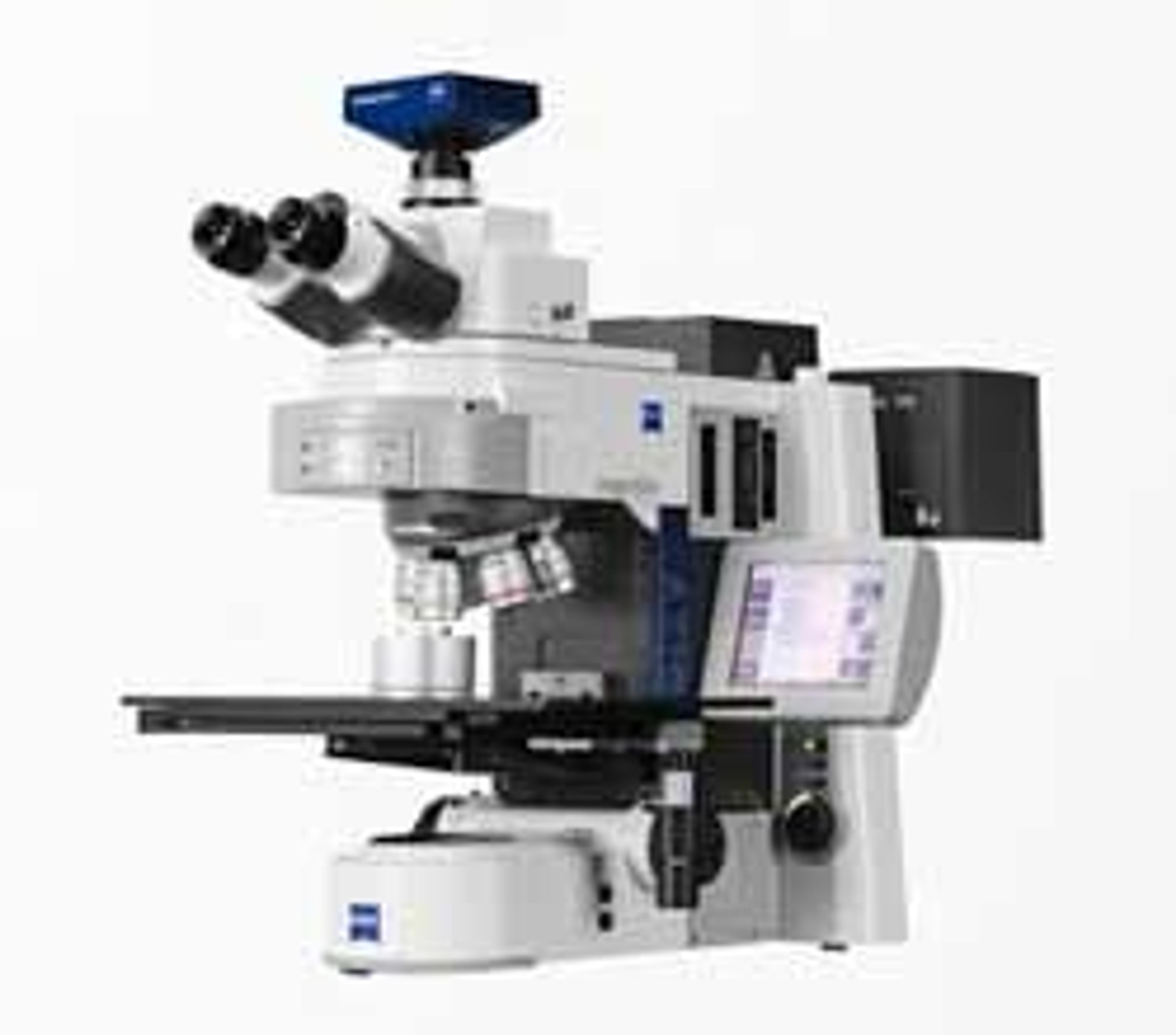
ZEISS Axio Imager 2 for Materials
ZEISS Research Microscopy SolutionsYour Open Microscope System for Automates Material Analysis.