Measuring Live Cells and Dead Cells in Real Time for Days Using a Plate Reader: Subsequent Multiplexing to Improve Efficiency and Reproducibility
17 Aug 2016This Scientific Poster demonstrates how Promega’s recently developed assay technologies make it possible to use multi-well plate readers to measure the number of live or dead cells in culture in real time over a period of days. In addition to providing real time kinetic measurements that are valuable for assay development and characterization activities, multiplexing with other assays provides a time saving approach and statistical advantage inherent in taking measurements from the same sample of cells.
Related products
Request Quote for All Products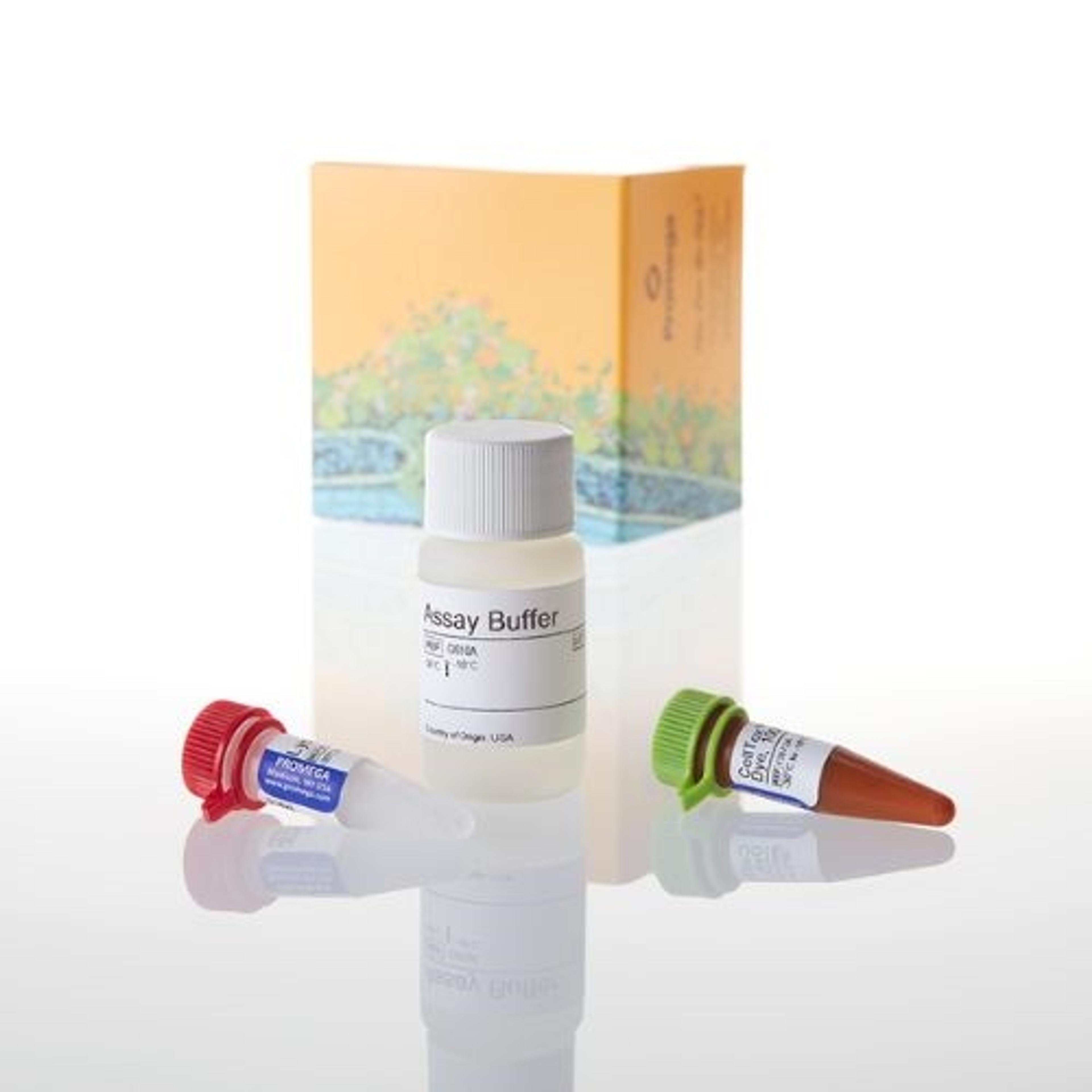
CellTox™ Green Cytotoxicity Assay
Promega Corp.The CellTox™ Green Cytotoxicity Assay measures changes in membrane integrity that occur as a result of cell death. The assay is intended to assess cytotoxicity in cell culture after experimental manipulation. The assay system uses a proprietry asymmetric cyanine dye that is excluded from viable cells but preferentially stains the DNA from dead cells. When the dye binds DNA released from cells, its fluorescence properties are substantially enhanced. Viable cells produce no appreciable increases in fluorescence. Therefore, the fluorescence signal produced by the binding interaction with dead cell DNA is proportional to cytotoxicity. The CellTox™ Green Dye is non-toxic to cells, and the signal remains constant after exposure of 72 hours, making it ideal for determining toxic effects of treatments throughout an extended exposure or as an endpoint determination. Features - Benefits • Accurate Cytotoxicity Determination: The CellTox™ Green Dye stably binds DNA of cells that have lost membrane integrity throughout 72-hour exposure and won’t underestimate cytotoxicity. • Kinetic Cytotoxicity Measures: Measure cytotoxicity at convenient time points from the same sample well to detect onset of toxicity with no duplication of plates. • Simple and Flexible Protocols: Add assay reagent directly to cells prior to plating or with dosing media to perform kinetic cytotoxicity measurements, eliminating a reagent dispensing step, or add diluted dye directly to cell culture wells as an endpoint add-mix-measure assay. • Multiplexing-Compatible: Get more informative data per well and reduce cell culture expenses by multiplexing with fluorescent and luminescent cell-based assays in the same well with no sample manipulation. • Easily Automated: Easily scale from 96- to 1536-well plate formats with “no-addition” or “single-addition” protocols. Applications Determine cytotoxic effects of treatments on cells in culture after long-term exposure.

CellTiter-Glo® Luminescent Cell Viability Assay
Promega Corp.The CellTiter-Glo® Luminescent Cell Viability Assay(a,b) is a homogeneous method of determining the number of viable cells in culture based on quantitation of the ATP present, an indicator of metabolically active cells. The CellTiter-Glo® Assay is designed for use with multiwell formats, making it ideal for automated high-throughput screening (HTS), cell proliferation and cytotoxicity assays. The homogeneous assay procedure involves adding the single reagent (CellTiter-Glo® Reagent) directly to cells cultured in serum-supplemented medium. Cell washing, removal of medium and multiple pipetting steps are not required. The system detects as few as 15 cells/well in a 384-well format in 10 minutes after adding reagent and mixing. The homogeneous "add-mix-measure" format results in cell lysis and generation of a luminescent signal proportional to the amount of ATP present. The amount of ATP is directly proportional to the number of cells present in culture. The CellTiter-Glo® Assay generates a "glow-type" luminescent signal, which has a half-life generally greater than five hours, depending on cell type and medium used. The extended half-life eliminates the need to use reagent injectors and provides flexibility for continuous or batch mode processing of multiple plates. The unique homogeneous format avoids errors that may be introduced by other ATP measurement methods that require multiple steps.