ResourceLife Sciences
Long-term live cell visualization and quantification of neuronal activity
25 Aug 2020In this application note, Sartorius presents data to support the use of the the IncuCyte S3 for Neuroscience to characterize and refine different neuronal phenotypes and model their function in vitro. Here, Sartorius explains how this single live-cell imaging platform allows users to assess calcium flux kinetics and continuously monitor the morphology of neuronal populations, using non-perturbing reagents, validated protocols that are cell-sparing, and a built-in, guided interface for non-experts, as provided by the IncuCyte S3 Neuronal Activity Analysis Software Module.
Related products
Request Quote for All Products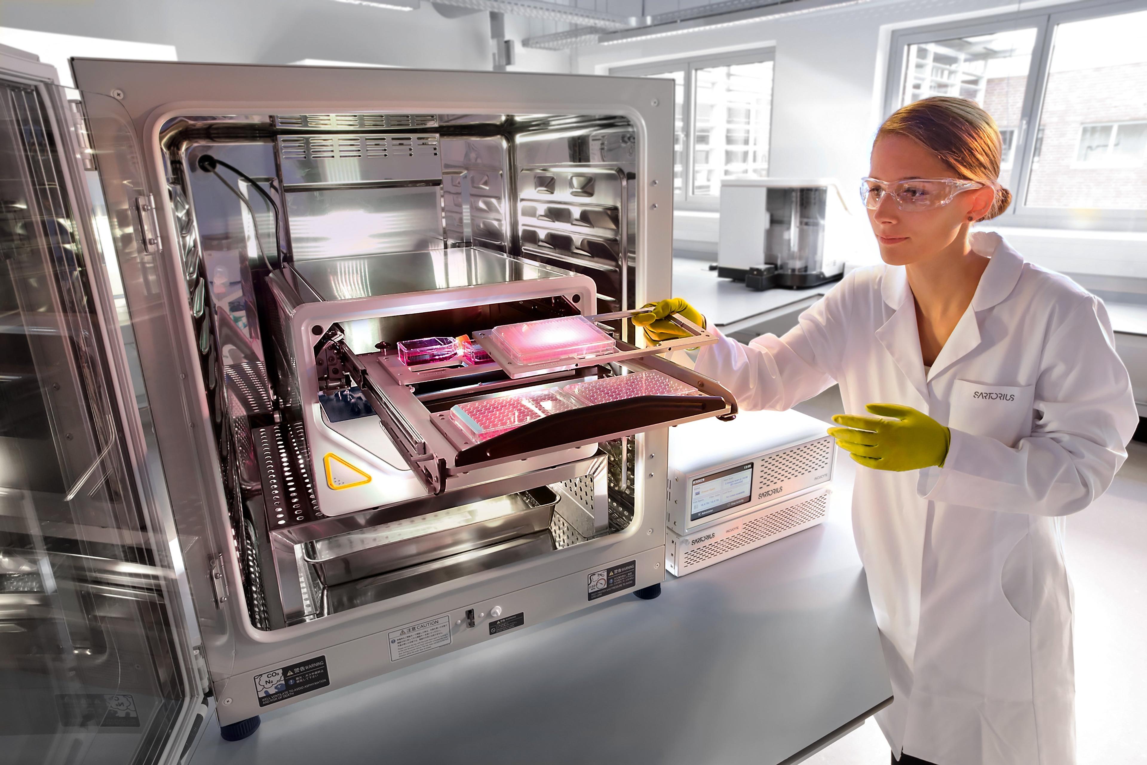
Incucyte® S3 Live-Cell Analysis System
Sartorius GroupOur flagship, supports the workflow and workload of larger laboratories.
Links
Tags
Cell / Tissue CultureCell culture or tissue culture is used to study the biology of cells or tissues and to isolate cellular products in an environment which can be manipulated and well defined. Accurately control your culture environment with bioreactors or culture incubators, bind your cells to a surface or together with an extracellular matrix. Distinguish cell types with differential media or proliferate cells with certain characteristics using selective media. Enrich your media with supplements such as growth factors, sera and vitamins. Find the best cell and tissue culture products, kits and equipment in our peer-reviewed product directory: compare products, check customer reviews and receive pricing direct from manufacturers.Gel Doc / Image AnalysisGel documentation (gel doc) or gel imaging systems are used for the analysis of proteins, antibodies and nucleic acid immobilized in polyacrylamide or agarose gels, membranes or microarrays. Explore a range of a gel imaging systems, densitometers, scanners, transilluminators or UV lamp + CCD cameras for your image analysis solutions. Colorimetric, fluorescent and/or radioisotopic samples can be visualized and documented for further analysis. See gel doc / Image analysis software for quantitative 1D and 2D analysis of your samples. Find the best gel doc / image analysis products in our peer-reviewed product directory: compare products, check customer reviews and receive pricing direct from manufacturers.In Vivo Imaging Systems<i>In vivo</i> imaging systems, including pre-clinical imaging systems and medical imaging systems are used to non-invasively visualize and capture images of live animals and plants. Monitor the natural processes or diseases of your subjects using small-animal pre-clinical imaging systems, including single photon positron emission tomography (SPECT), positron emission tomography (PET), computed tomography (micro-CT), magnetic resonance imaging (MRI), X-ray radiography, ultrasound, fluorescence and bioluminescence imagers. Multimodal systems and software solutions are also available for correlative analysis of organ, tissue, cell, or molecular-level processes. Find the best in vivo imaging products in our peer-reviewed product directory: compare products, check customer reviews and receive pricing direct from manufacturers.NeuroscienceNeuroscience research investigates the neurological mechanisms underlying behavior, neurodegenerative diseases, and other brain conditions. Learn about the innovative technologies for bioimaging, electrophysiology, cell culture, chromatography and other techniques used in this field.Live Cell ImagingLive cell imaging is the study of living cells using microscopes and high-content imaging systems. This technique provides in-depth insight into fast and complex biological processes, by allowing dynamic imaging of living cells instead of acquiring an individual image at a single point in time.