Investigating structure-property relationships in a carbon-fiber composite
19 Jun 2024Characterizing composite materials is a challenging task. Understanding the nucleation processes is critical toward engineering against failure, but traditional bulk testing methods are insufficient to describe this process. ZEISS presents how correlative microscopy is a viable conduit into the digital material testing approach. A carbon fiber reinforced composite hockey stick was used as the subject of the characterization study, though this same technique can apply to any variety of materials, from glass composites to metal matrix composites, as well as to monolithic materials. Correlative microscopy enabled a robust imaging-to-simulation workflow, producing a model that is available for further digital modification and analysis. Through implementation of this procedure in a regular basis, material development efficiencies may be enhanced, leading to high-performance products in a reduced amount of time.
Related products
Request Quote for All Products
ZEISS Atlas 5
ZEISS Research Microscopy SolutionsLarge area imaging for SEM, FE-SEM & FIB-SEM ATLAS combines a 16 bit scan generator and dual super-sampling signal acquisition hardware with image processing and control software for your ZEISS electron microscope. Acquire images up to 32 k x 32 k pixels, with dwell times from 100 ns to > 100 s, adjustable in 100 ns increments. Save your images with eight or sixteen bits of intensity. With the ATLAS “Mosaic Tool” you create large image montages, automatically moving from image tile to tile, and mosaic site to site, resulting in an “Extreme Field of View” image, at SEM nanometer scale resolution. ATLAS provides • reduced number of tiles to acquire, reducing stage motion delay and areal fraction of each image “lost” to overlap • reduced number of overlap “seams”, leading to less beam damage and degradation of the sample • reduced computational complexity
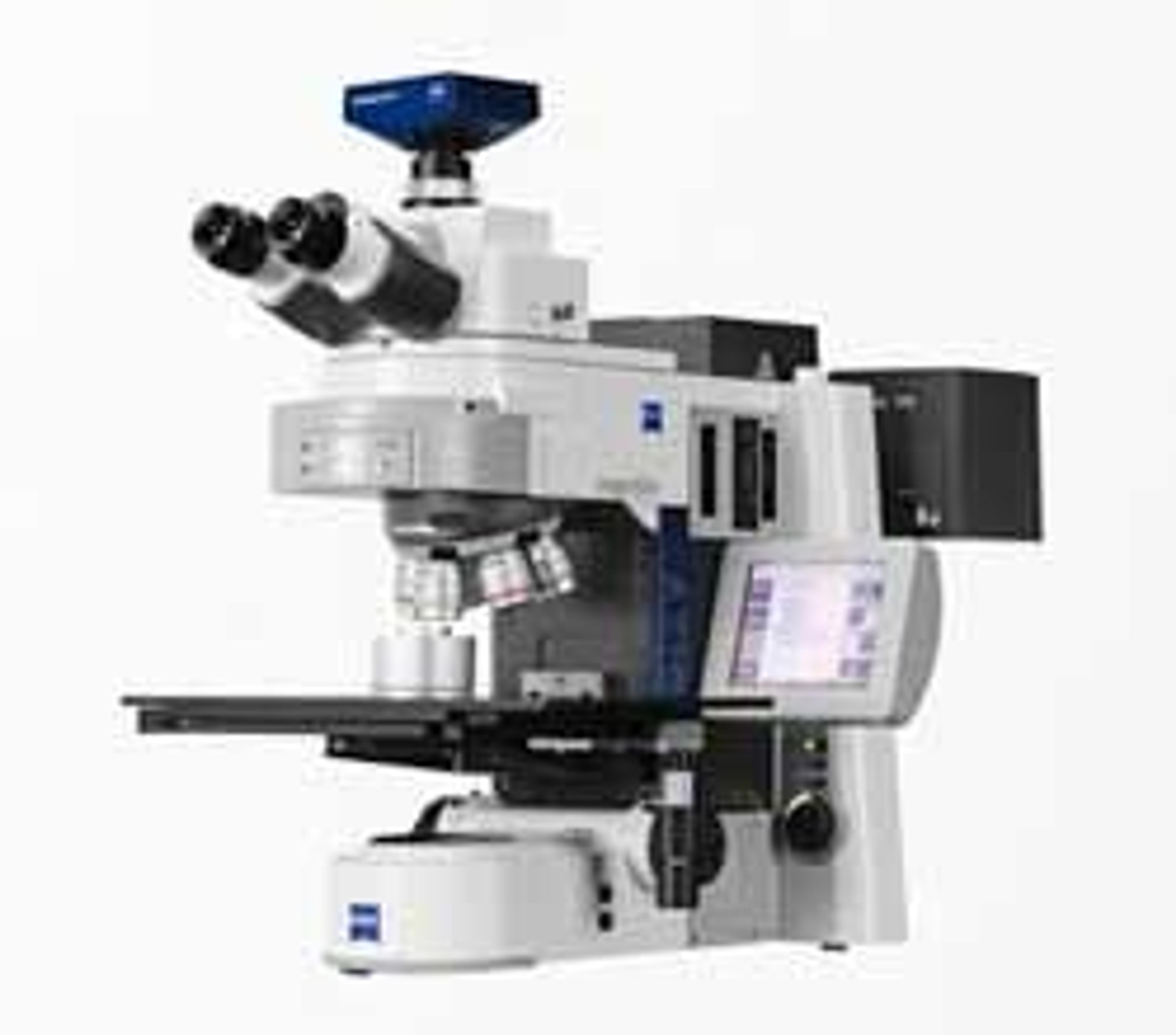
ZEISS Axio Imager 2 for Materials
ZEISS Research Microscopy SolutionsYour Open Microscope System for Automates Material Analysis.

ZEISS Xradia Versa X-ray Microscopes
ZEISS Research Microscopy SolutionsDiscover More with Non-destructive 3D X-ray Imaging at Submicron Resolution