ResourceLife Sciences
In vivo imaging with GFP: From blue squeezates to green mice
10 Nov 2025In this application note, Analytik Jena shows how the UVP iBox Studio can be used to perform in vivo imaging of an orthotopic mouse model of pancreatic cancer labeled with GFP. The experiment highlights not only the striking detection capability of the instrument to visualize a deep tumor, but the fine detail that can be captured with its high sensitivity, deeply cooled, low noise detector.
Related products
Request Quote for All Products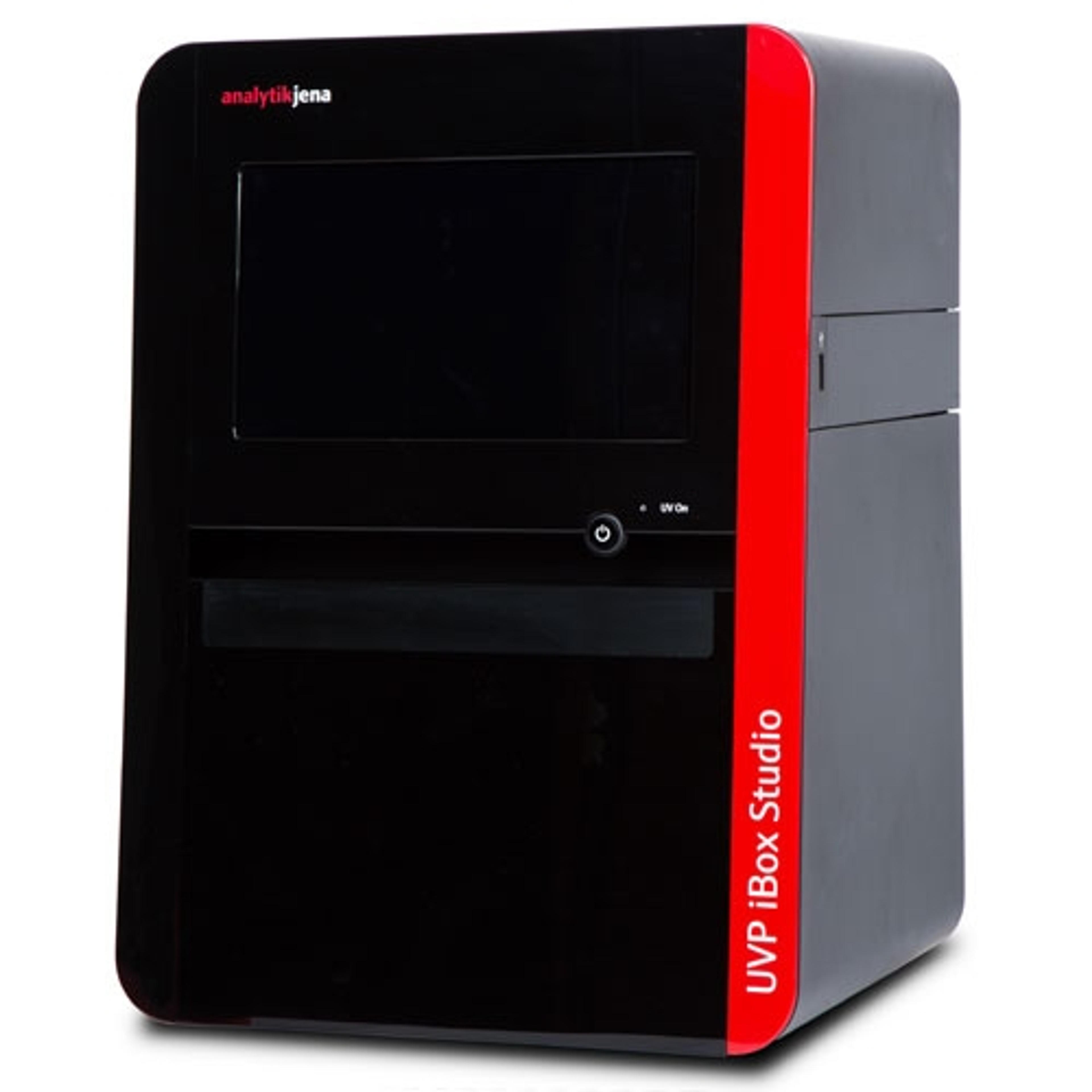
UVP iBox Studio touch
UVP, An Analytik Jena CompanyFor researchers with limited focus on, or just starting in vivo pre-clinical studies, the UVP iBox Studio provides highly sensitive fluorescence in vivo imaging at an affordable price.
Links
Tags
In Vivo Imaging Systems<i>In vivo</i> imaging systems, including pre-clinical imaging systems and medical imaging systems are used to non-invasively visualize and capture images of live animals and plants. Monitor the natural processes or diseases of your subjects using small-animal pre-clinical imaging systems, including single photon positron emission tomography (SPECT), positron emission tomography (PET), computed tomography (micro-CT), magnetic resonance imaging (MRI), X-ray radiography, ultrasound, fluorescence and bioluminescence imagers. Multimodal systems and software solutions are also available for correlative analysis of organ, tissue, cell, or molecular-level processes. Find the best in vivo imaging products in our peer-reviewed product directory: compare products, check customer reviews and receive pricing direct from manufacturers.GFP