Imaging living cells without compromising cell integrity

28 Aug 2019

The HoloMonitor® label-free live cell imaging system is based on the principle of quantitative phase imaging, enabling non-invasive visualization and quantification of living cells without compromising cell integrity. In this application note, Phase Holographic Imaging describes the rationale and advantages of using quantitative phase imaging for live cell kinetic analysis of cellular events — explaining the power of live cell time-lapse cytometry and how cells are made visible without labels or stains.

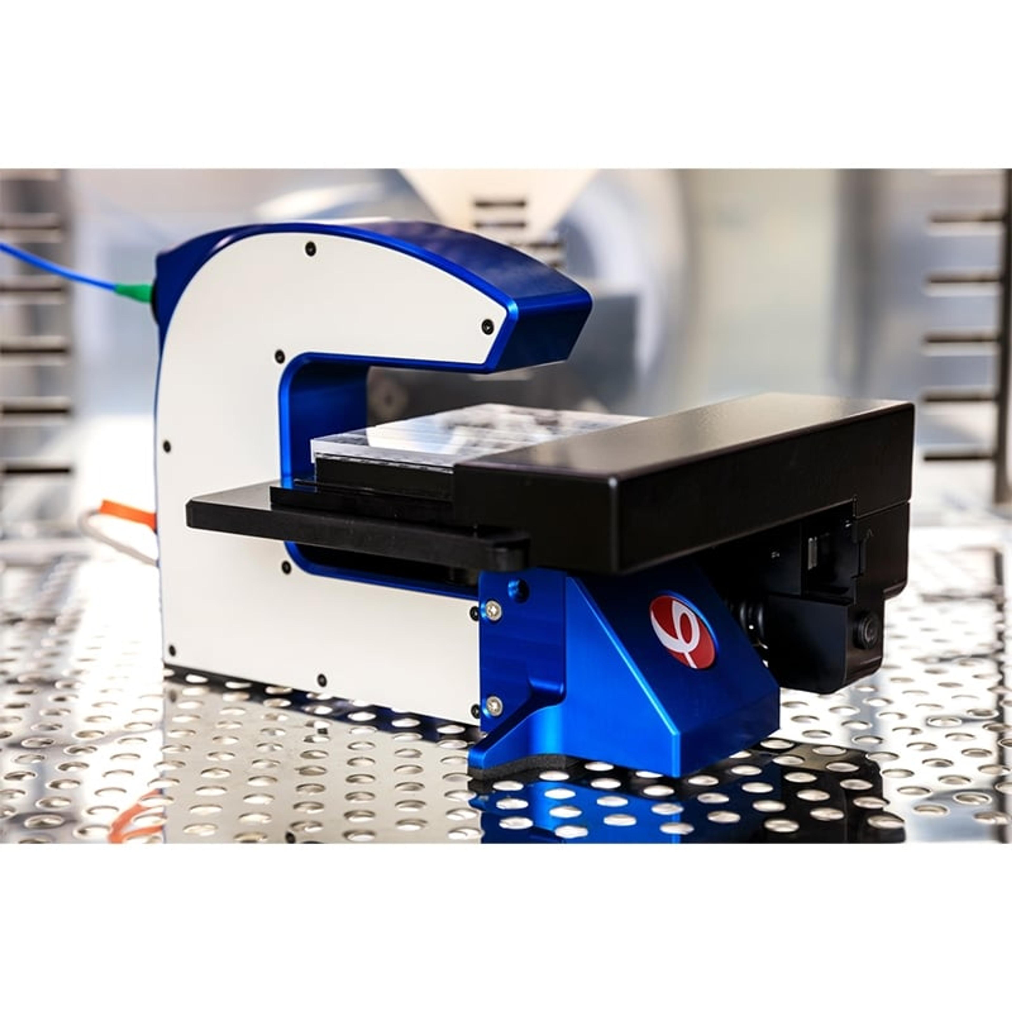
HoloMonitor® M4 Live Cell Analysis System

Phase Holographic Imaging (PHI)

The HoloMonitor ® live cell imaging system enables long-term non-invasive analysis of cell cultures within your standard incubator. Cell biologists worldwide use PHI’s label-free cell imager to automatically obtain accurate live-cell morphology, migration, and proliferation data as well as multi-well time-lapse videos, down to a single-cell level in real-time.

(19)

Links

Tags

Cell / Tissue CultureCell culture or tissue culture is used to study the biology of cells or tissues and to isolate cellular products in an environment which can be manipulated and well defined. Accurately control your culture environment with bioreactors or culture incubators, bind your cells to a surface or together with an extracellular matrix. Distinguish cell types with differential media or proliferate cells with certain characteristics using selective media. Enrich your media with supplements such as growth factors, sera and vitamins. Find the best cell and tissue culture products, kits and equipment in our peer-reviewed product directory: compare products, check customer reviews and receive pricing direct from manufacturers.Flow Cytometry / Cell CountingFlow cytometers are used to count, sort and examine multiple characteristics of cells. Other cell analysis equipment includes image cytometers, cell counters, fluorescence-activated cell sorters (FACS), magnetic-activated cell sorters (MACS), and a range of flow cytometry assay kits. Flow cytometers can reveal information on cell viability, cell proliferation, apoptosis and cell cycle progression, as well as identify cell populations and intracellular or cell-surface molecules. Additionally, some flow cytometers, known as FACS, have an additional sorting function after analysis. Cell counters and image cytometers count live and dead cell populations and can also conduct cell proliferation assays. Find the best flow cytometers, cell counters and cell sorters in our peer-reviewed product directory: compare products, check customer reviews and receive pricing direct from manufacturers.CytometryCytometry is a broad term for the quantitative analysis of cells and cell systems. The most popular methods to study cytometry are flow cytometry and image cytometry.Cell ImagingCell imaging can be achieved using a number of techniques including confocal microscopy, transmission electron microscopy, atomic force microscopy, and light sheet microscopy.Live Cell ImagingLive cell imaging is the study of living cells using microscopes and high-content imaging systems. This technique provides in-depth insight into fast and complex biological processes, by allowing dynamic imaging of living cells instead of acquiring an individual image at a single point in time.Non-Invasive ImagingLabel-Free Analysis
Imaging living cells without compromising cell integrity