Imaging breast cancer evolution in 3D organoid cultures with Luxendo light-sheet microscopy
17 Jul 2023Bruker's Luxendo light-sheet technology can image a wide range of live, fixed, and cleared biological samples from organoids and embryos to large whole organisms. Overcoming many of the barriers that conventional light microscopy faces, lightsheet fluorescence microscopy, also called selective plane illumination microscopy (SPIM), supports advanced research in the life sciences. SPIM works by de-coupling the fluorescence excitation and detection beams and together with a sheet of light for excitation, selectively illuminates a focal plane for high-resolution images without the danger of phototoxicity. Here, Dr. Martin Jechlinger, the Senior Scientist and Head of VISION Laboratory at the MOLIT Institute, talks about how his lab utilizes light-sheet microscopy to investigate 3D organoid cultures. Specifically, the molecular mechanisms underlying mammary tumor development during breast cancer, as well as what is happening when treatments are failing.
Related products
Request Quote for All Products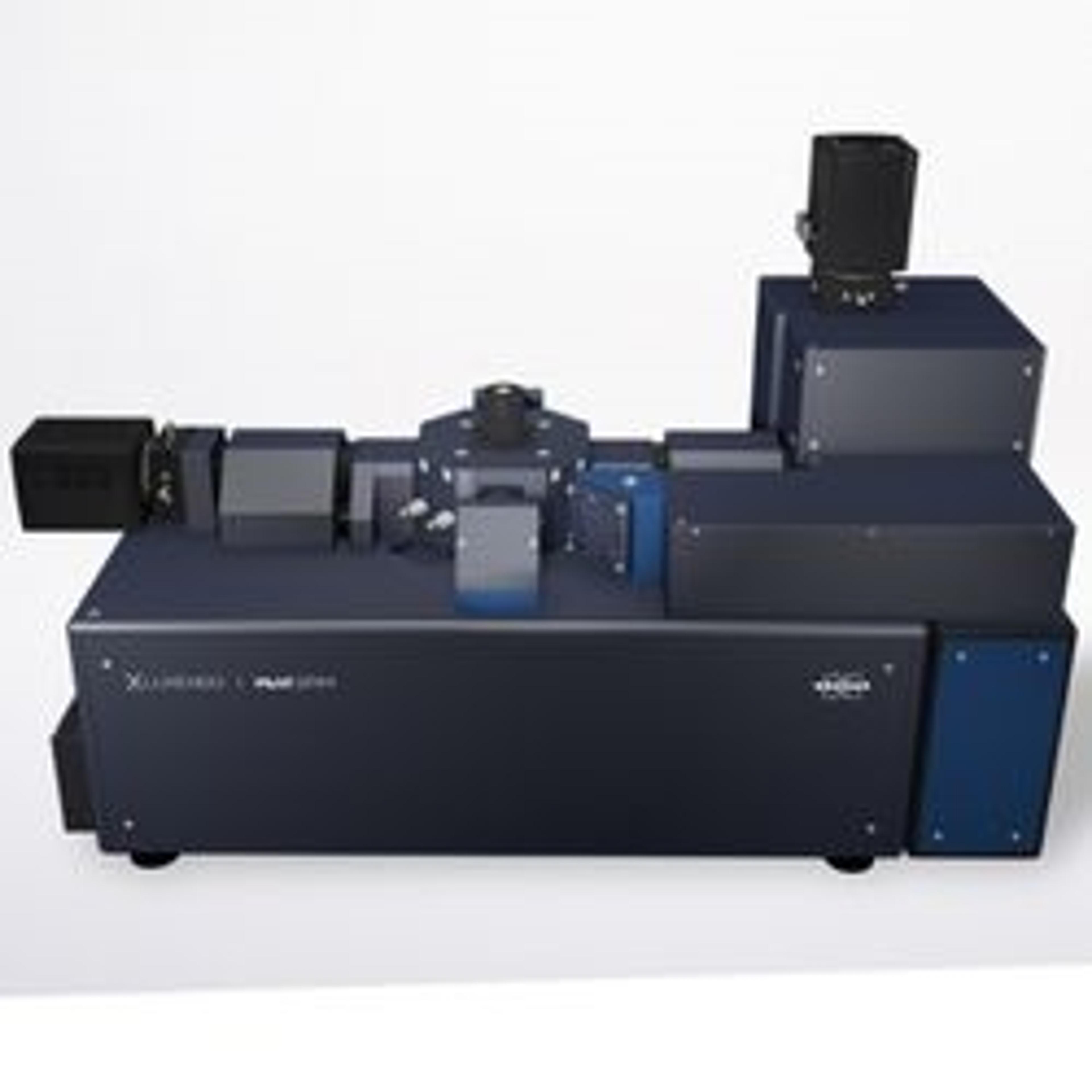
MuVI SPIM
Bruker Fluorescence MicroscopyBruker's MuVi Selective-Plane Illumination Microscope (SPIM) is the fastest multi-angle view system on the market. MuVi incorporates years of Luxendo light-sheet technology innovations to provide high-speed volumetric acquisition for analyzing dynamic processes in delicate live specimens, as well as offering compatibility with any clearing method. A variety of other advanced hardware and software integrations can be easily configured to support evolving multidimensional experimental needs.