ResourceLife Sciences
How to Improve Your Fluorescent Western Blots
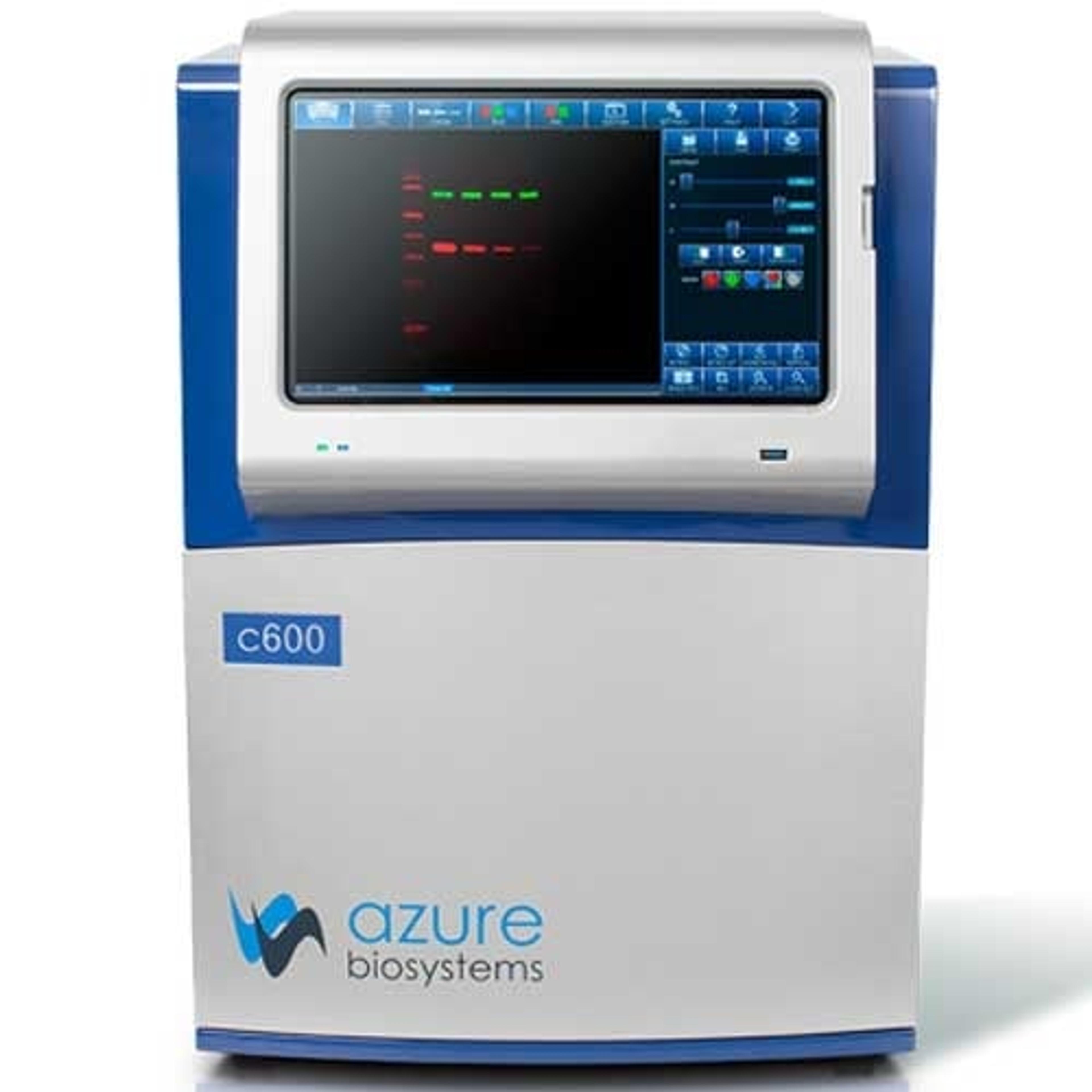
4 Feb 2015Fluorescent Western blots are the gold standard for quantitative Westerns. They are ideal for detecting multiple proteins simultaneously (multiplexing), allowing in-lane normalization and detection of your protein of interest and loading control at the same time. In addition, post-translational modifications can be studied and quantitated easily. This application note suggests how to improve your fluorescent Western blots.
Related products
Request Quote for All Products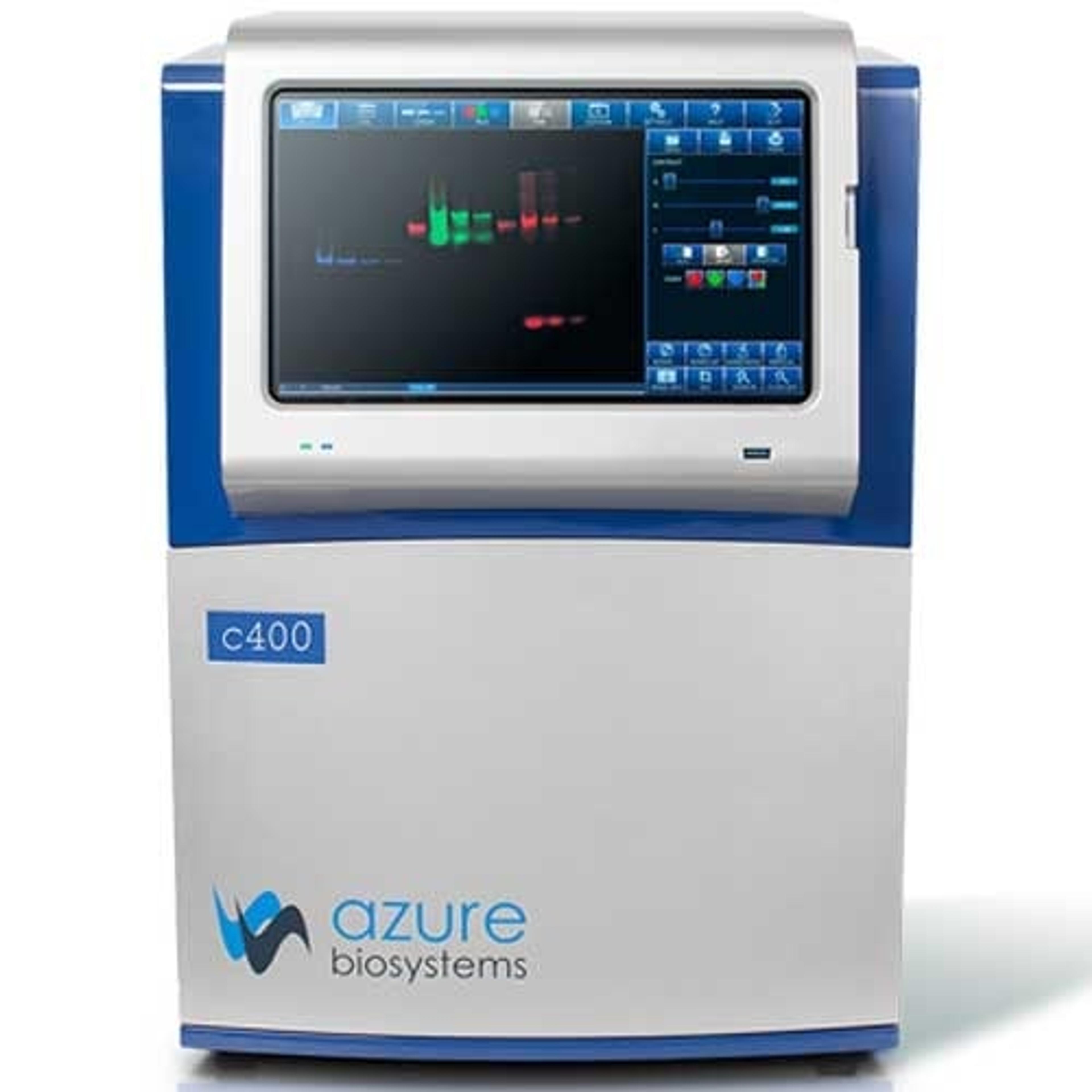

Links
Tags
AntibodiesAntibodies are used in techniques such as confocal and fluorescence microscopy, flow cytometry, ELISA, ELISPOT, immunohistochemistry, western blotting and immunopreciptation. Select specific antigen reactivity, high specific affinity, low non-specific binding, monoclonal or polyclonal, primary or secondary antibodies and associated conjugates such as an enzyme or dye for visualization.Western BlottingWestern blotting equipment is used to transfer and identify specific proteins within a sample, reveal protein modifications, as well as give a semi-quantitative estimation of their concentration. Western blotting equipment includes all apparatus necessary to transfer proteins from gel to membrane and subsequent processing steps. Protein transfer can be performed by electroblotting with wet, semi-dry and dry transfer systems onto nitrocellulose and PVDF membranes. Blocking, washing and labeling of membranes follows, involving buffers, blocking reagents, blotting / incubation trays, labeling reagents, immunoblotting assays, antibodies and conjugates. Automated equipment for these steps is available to accelerate your lab workflow. Finally, detection and imaging of proteins can be conducted using gel documentation and imaging systems. Find the best western blotting equipment in our peer-reviewed product directory: compare products, check customer reviews and receive pricing direct from manufacturers.Gel Doc / Image AnalysisGel documentation (gel doc) or gel imaging systems are used for the analysis of proteins, antibodies and nucleic acid immobilized in polyacrylamide or agarose gels, membranes or microarrays. Explore a range of a gel imaging systems, densitometers, scanners, transilluminators or UV lamp + CCD cameras for your image analysis solutions. Colorimetric, fluorescent and/or radioisotopic samples can be visualized and documented for further analysis. See gel doc / Image analysis software for quantitative 1D and 2D analysis of your samples. Find the best gel doc / image analysis products in our peer-reviewed product directory: compare products, check customer reviews and receive pricing direct from manufacturers.Protein PurificationProtein purification is a vital step in drug discovery, therapeutics, biotech and life science research. The purification process typically involves subcellular or membrane protein extraction with cell lysis kits, separation of proteins from cell debris by filtration or spin columns, and the isolation of proteins of interest from other proteins and impurities with affinity purification (including fusion protein tags and antibody binding proteins A, G and L), immunoprecipitation or chromatographic methods, such as ion exchange, size exclusion and immobilized metal affinity chromatography. All purification methods come in multiple formats for your laboratory needs, including agarose or magnetic beads, resins, columns and filter plates. Find the best protein purification equipment in our peer-reviewed product directory: compare products, check customer reviews and receive pricing direct from manufacturers.Method DevelopmentMethod development is the process of creating and optimizing experimental techniques and protocols to achieve reliable, reproducible results. This is essential in various fields, including pharmaceuticals, environmental science, and diagnostics. Browse our peer-reviewed product directory to find the best method development solutions, compare products, check reviews, and get pricing directly from manufacturers.Blotting