HoloMonitor M4 product brochure

20 Aug 2023

In this product brochure, Phase Holographic Imaging (PHI) presents the HoloMonitor® M4 live cell imaging system, offering non-invasive, long-term analysis of cell cultures within standard incubators. The system enables the automatic capture of label-free live-cell morphology, migration, and proliferation data in real-time, down to a single-cell level. HoloMonitor is designed for the visualization and quantification of adherent cells over time, suitable for various cell types, including delicate ones like induced pluripotent stem cells and primary human cell cultures. The label-free imaging preserves cell viability for subsequent studies. The HoloMonitor software is a user-friendly tool that extracts quantitative data from holographic images, guiding users through experiment setup and result analysis. The software also allows easy creation of visual content for publications, and data can be exported to Excel for further analysis.

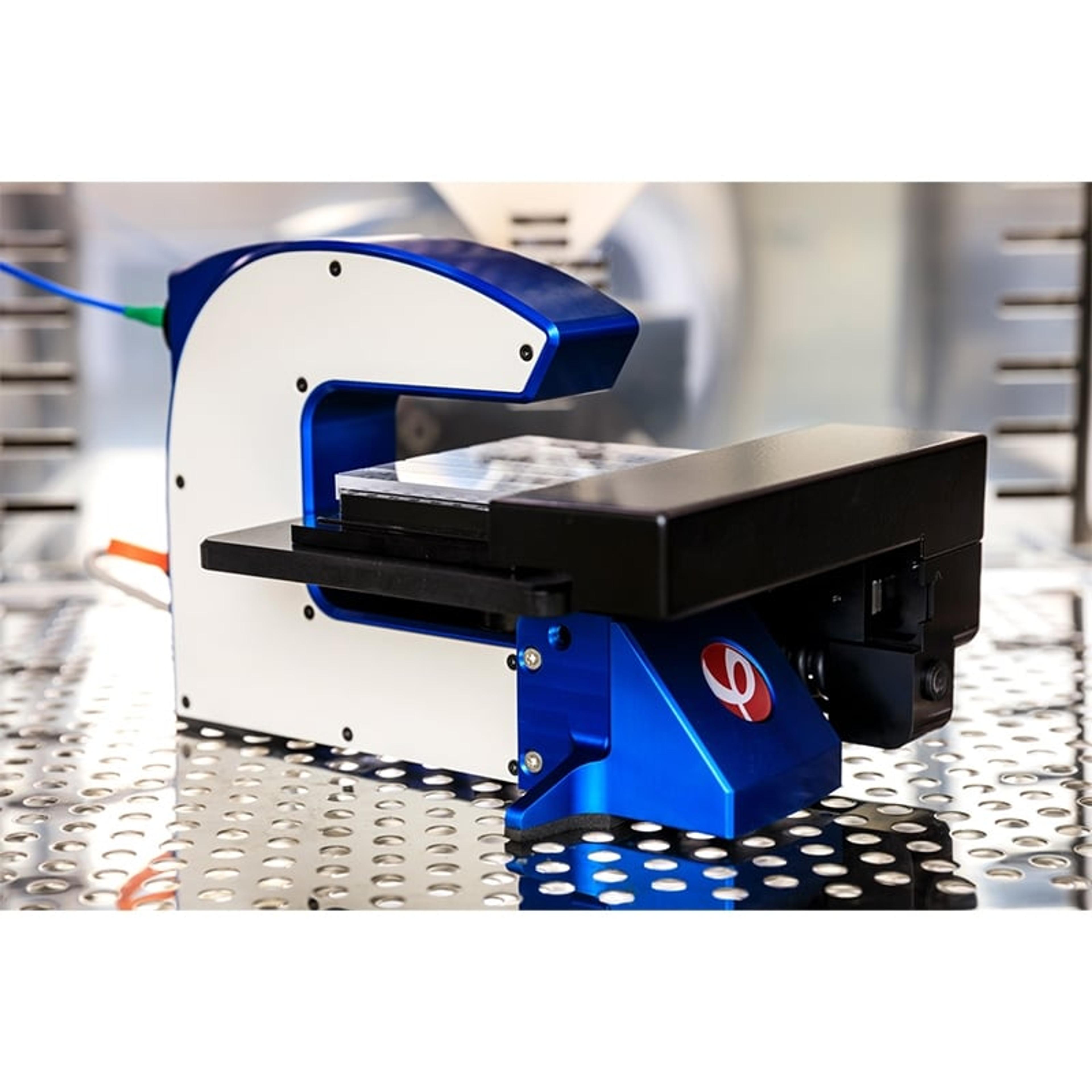
HoloMonitor® M4 Live Cell Analysis System

Phase Holographic Imaging (PHI)

The HoloMonitor ® live cell imaging system enables long-term non-invasive analysis of cell cultures within your standard incubator. Cell biologists worldwide use PHI’s label-free cell imager to automatically obtain accurate live-cell morphology, migration, and proliferation data as well as multi-well time-lapse videos, down to a single-cell level in real-time.

(19)

Links

Tags

In Vivo Imaging Systems<i>In vivo</i> imaging systems, including pre-clinical imaging systems and medical imaging systems are used to non-invasively visualize and capture images of live animals and plants. Monitor the natural processes or diseases of your subjects using small-animal pre-clinical imaging systems, including single photon positron emission tomography (SPECT), positron emission tomography (PET), computed tomography (micro-CT), magnetic resonance imaging (MRI), X-ray radiography, ultrasound, fluorescence and bioluminescence imagers. Multimodal systems and software solutions are also available for correlative analysis of organ, tissue, cell, or molecular-level processes. Find the best in vivo imaging products in our peer-reviewed product directory: compare products, check customer reviews and receive pricing direct from manufacturers.Light MicroscopyLight microscopes or optical microscopes are used to visualize microscale objects under magnification, including cells, clinical specimens and materials. Lab equipment for light microscopy includes confocal microscopes, fluorescence microscopes, zoom and stereo microscopes. Microscope slides and imaging reagents are available for visualizing samples, as well as various microscope stages and incubators for large or temperature-sensitive samples. Find the best light microscopes in our peer-reviewed product directory: compare products, check customer reviews and receive pricing direct from manufacturers.Cell ImagingCell imaging can be achieved using a number of techniques including confocal microscopy, transmission electron microscopy, atomic force microscopy, and light sheet microscopy.Cell AnalysisThe analysis of cells allows researchers to understand the factors which contribute to cell health and function. These influencing processes can then be predicted and altered, leading to the development of medication and disease treatments.Live Cell ImagingLive cell imaging is the study of living cells using microscopes and high-content imaging systems. This technique provides in-depth insight into fast and complex biological processes, by allowing dynamic imaging of living cells instead of acquiring an individual image at a single point in time.