ResourceLife Sciences
Fluorescent Image Analysis – Cell Division in Spheroids
6 Jun 2019Cell division inside spheroids can be visualized by staining cell nuclei and microtubules. The number of cells in mitosis in spheroids can be quantitatively evaluated using images captured by a laser confocal microscope and NoviSight™ software's counting module.
Related products
Request Quote for All Products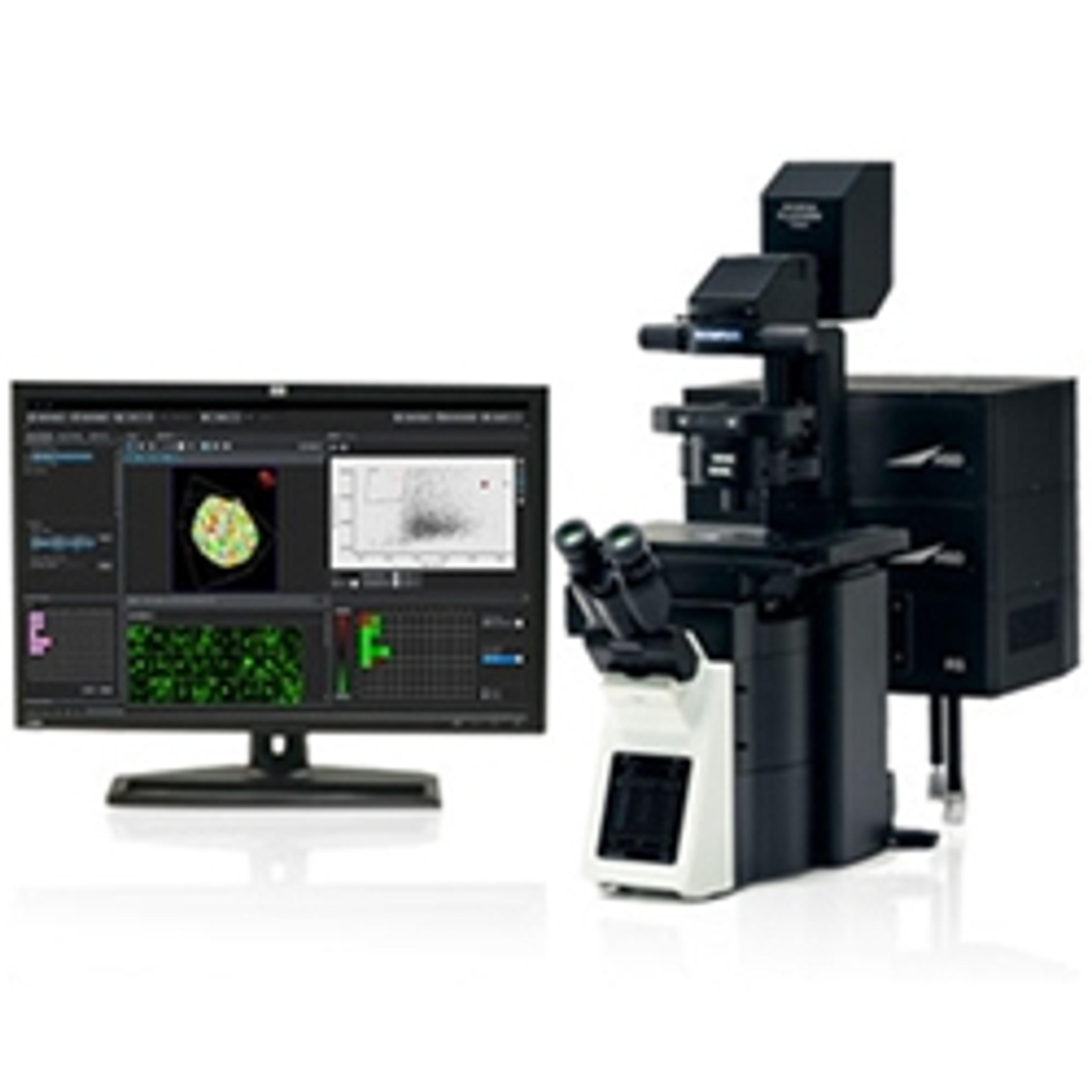
Links
Tags
Cell / Tissue CultureCell culture or tissue culture is used to study the biology of cells or tissues and to isolate cellular products in an environment which can be manipulated and well defined. Accurately control your culture environment with bioreactors or culture incubators, bind your cells to a surface or together with an extracellular matrix. Distinguish cell types with differential media or proliferate cells with certain characteristics using selective media. Enrich your media with supplements such as growth factors, sera and vitamins. Find the best cell and tissue culture products, kits and equipment in our peer-reviewed product directory: compare products, check customer reviews and receive pricing direct from manufacturers.FluorescenceThe emission of fluorescence occurs when a photon of energy is supplied to a fluorescent chemical compound by an external source, causing it to become excited. Fluorescence can be detected and measured for different purposes using microplate readers, fluorescence microscopes, fluorescence scanners, and flow cytometers.Cell AnalysisThe analysis of cells allows researchers to understand the factors which contribute to cell health and function. These influencing processes can then be predicted and altered, leading to the development of medication and disease treatments.Image AnalysisImage analysis involves the extraction of meaningful information from images, often using software to quantify and interpret visual data. It is widely used in cell biology, material science, and diagnostics. Increasingly, AI is being used to streamline image analysis. Explore image analysis tools in our peer-reviewed product directory; compare products, check reviews, and get pricing directly from manufacturers.Live Cell ImagingLive cell imaging is the study of living cells using microscopes and high-content imaging systems. This technique provides in-depth insight into fast and complex biological processes, by allowing dynamic imaging of living cells instead of acquiring an individual image at a single point in time.SpheroidsSpheroids are clusters of cells that have been grown in 3D culture to be used as <i>in vitro</i> model systems. These 3D microtissues can be used for toxicology testing, DMPK studies and many other applications involving cell analysis.Cancer ResearchAlthough cancer is often referred to as a single condition, it actually consists of more than 100 different diseases. Microscopy, mass spectrometry, high throughput sequencing and flow cytometry are some of the most common techniques employed in cancer research labs.