ResourceLife Sciences
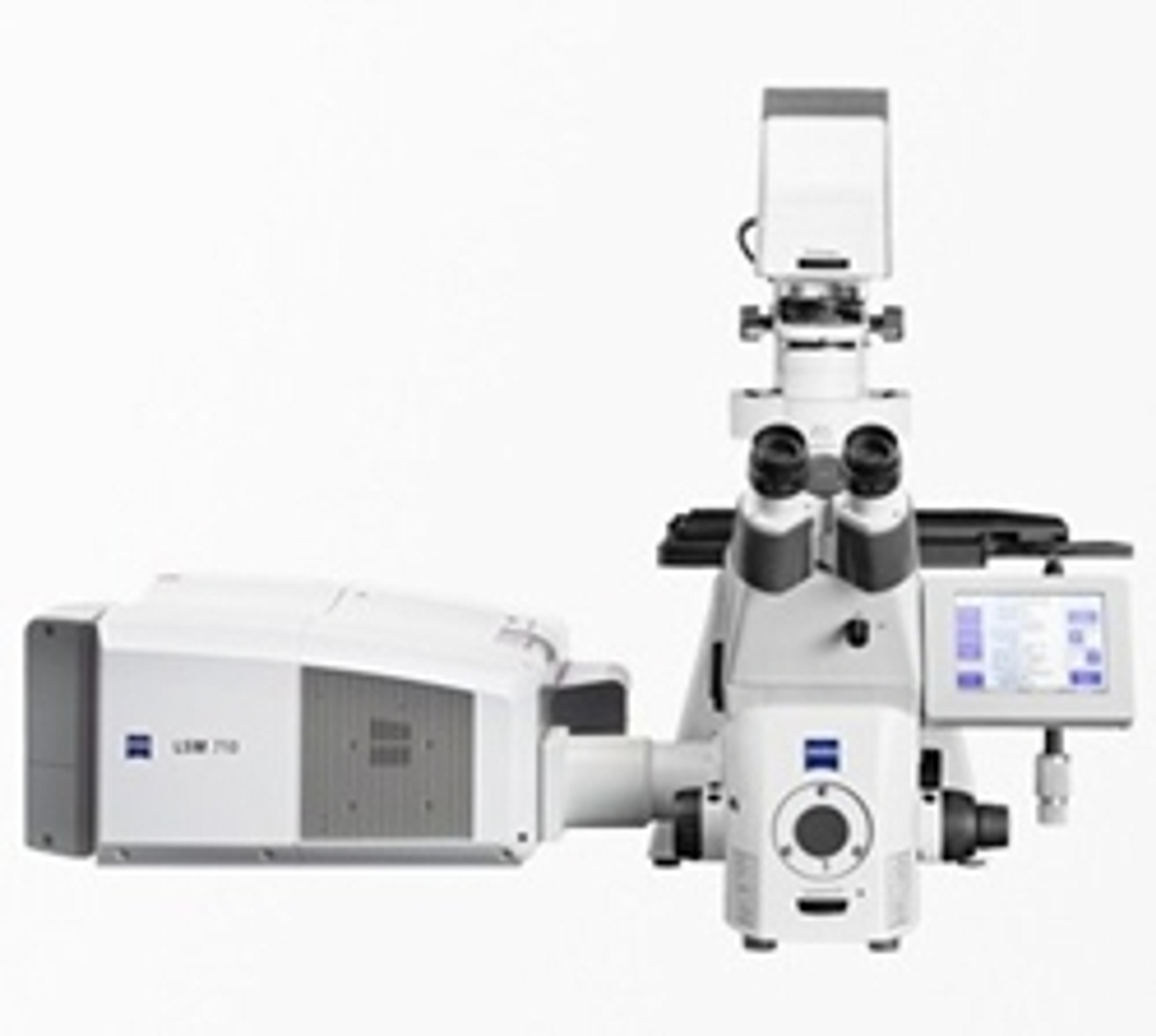
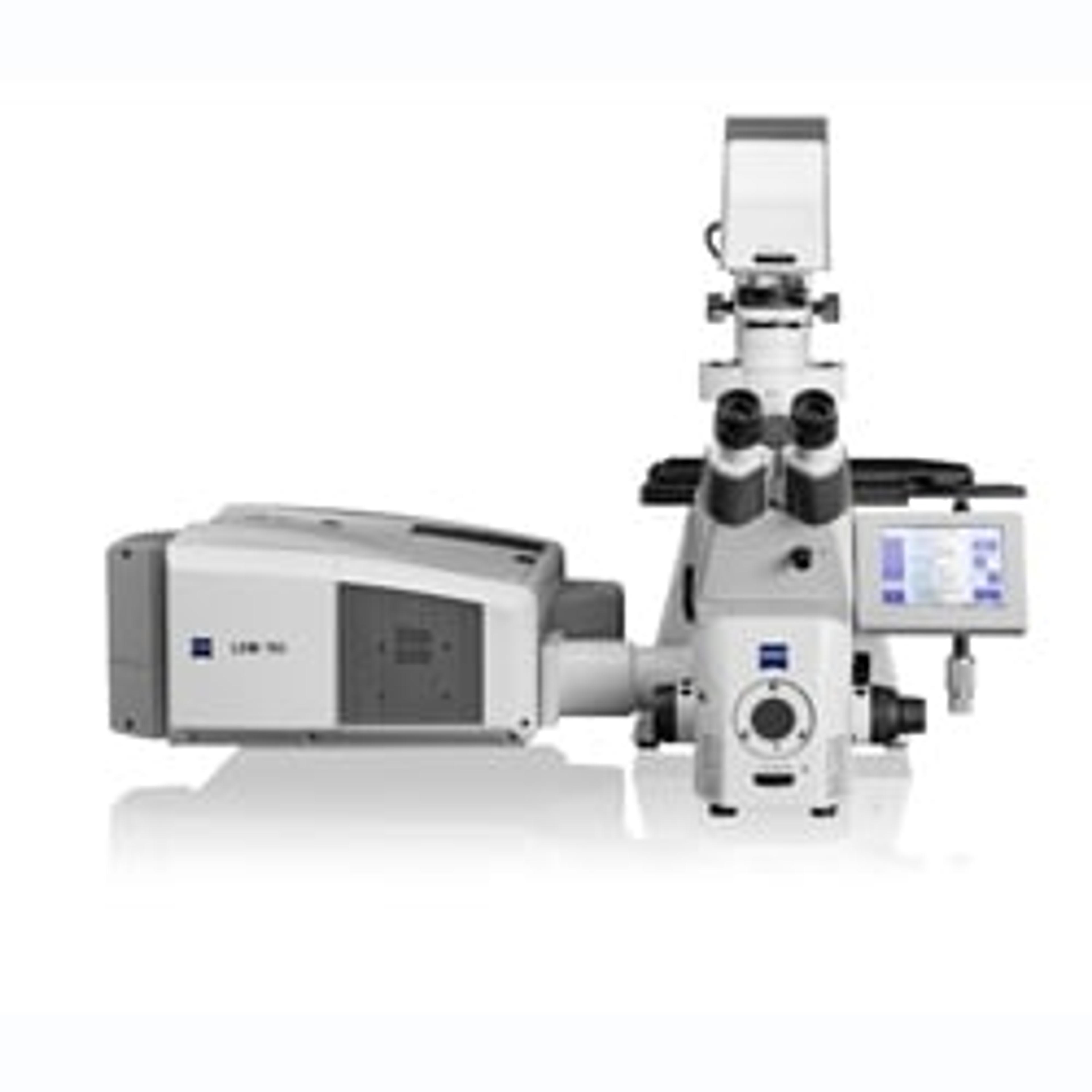
Fluorescence Polarization and Anisotropy Imaging with LSM 710/LSM 780
4 May 2018In this white paper, ZEISS describes a general method for the determination of instrumental correction factor G, for fluorescence polarization and anisotropy experiments utilizing an anisotropy standard.
Links
Tags
Light MicroscopyLight microscopes or optical microscopes are used to visualize microscale objects under magnification, including cells, clinical specimens and materials. Lab equipment for light microscopy includes confocal microscopes, fluorescence microscopes, zoom and stereo microscopes. Microscope slides and imaging reagents are available for visualizing samples, as well as various microscope stages and incubators for large or temperature-sensitive samples. Find the best light microscopes in our peer-reviewed product directory: compare products, check customer reviews and receive pricing direct from manufacturers.FluorescenceThe emission of fluorescence occurs when a photon of energy is supplied to a fluorescent chemical compound by an external source, causing it to become excited. Fluorescence can be detected and measured for different purposes using microplate readers, fluorescence microscopes, fluorescence scanners, and flow cytometers.Cell ImagingCell imaging can be achieved using a number of techniques including confocal microscopy, transmission electron microscopy, atomic force microscopy, and light sheet microscopy.ImagingImaging techniques are essential for obtaining visual representations of samples to understand structures, processes, and function in biological, chemical, and physical research. These tools range from traditional light microscopy to advanced imaging modalities like MRI and electron microscopy, providing researchers with valuable data for diagnostics, drug discovery, and material analysis. Explore imaging solutions in our peer-reviewed product directory to compare products, check reviews, and get pricing directly from manufacturers.