Considerations for cleared tissue imaging with light-sheet fluorescence microscopy in neuroscience research
18 Jul 2023A major goal in the field of neuroscience is to understand the connectivity of the brain across scales, from individual synaptic connections to whole organism connectomics. Visualizing the whole brain often requires imaging at depths that are not supported by conventional imaging methods, such as laser scanning confocal microscopy. In recent years, light-sheet fluorescence microscopy (LSFM) has emerged as an imaging technique capable of addressing a wide variety of applications in neuroscience due to its faster acquisition speed and larger imaging depth compared to confocal microscopy. Although early implementations of LSFM were optimized around imaging developmental processes in typical model organisms, technological advances in optics have allowed researchers to extend this technique to much larger samples, such as an entire mouse brain. Combined with emerging tissue clearing techniques, researchers are now able to investigate intact tissues, organs, and even entire organisms with subcellular resolution using light microscopy methods. The versatility of LSFM for neuroscience research has contributed to its rapid adoption and increasing popularity in the scientific community. However, there are multiple important factors one must consider when moving from standard imaging techniques to LSFM. In this application note, Bruker discusses the reasons why LSFM and clearing techniques are particularly useful for neuroscience research, important considerations for neuroscientists adopting this technology, and application examples of LSFM for cleared sample imaging.
Related products
Request Quote for All Products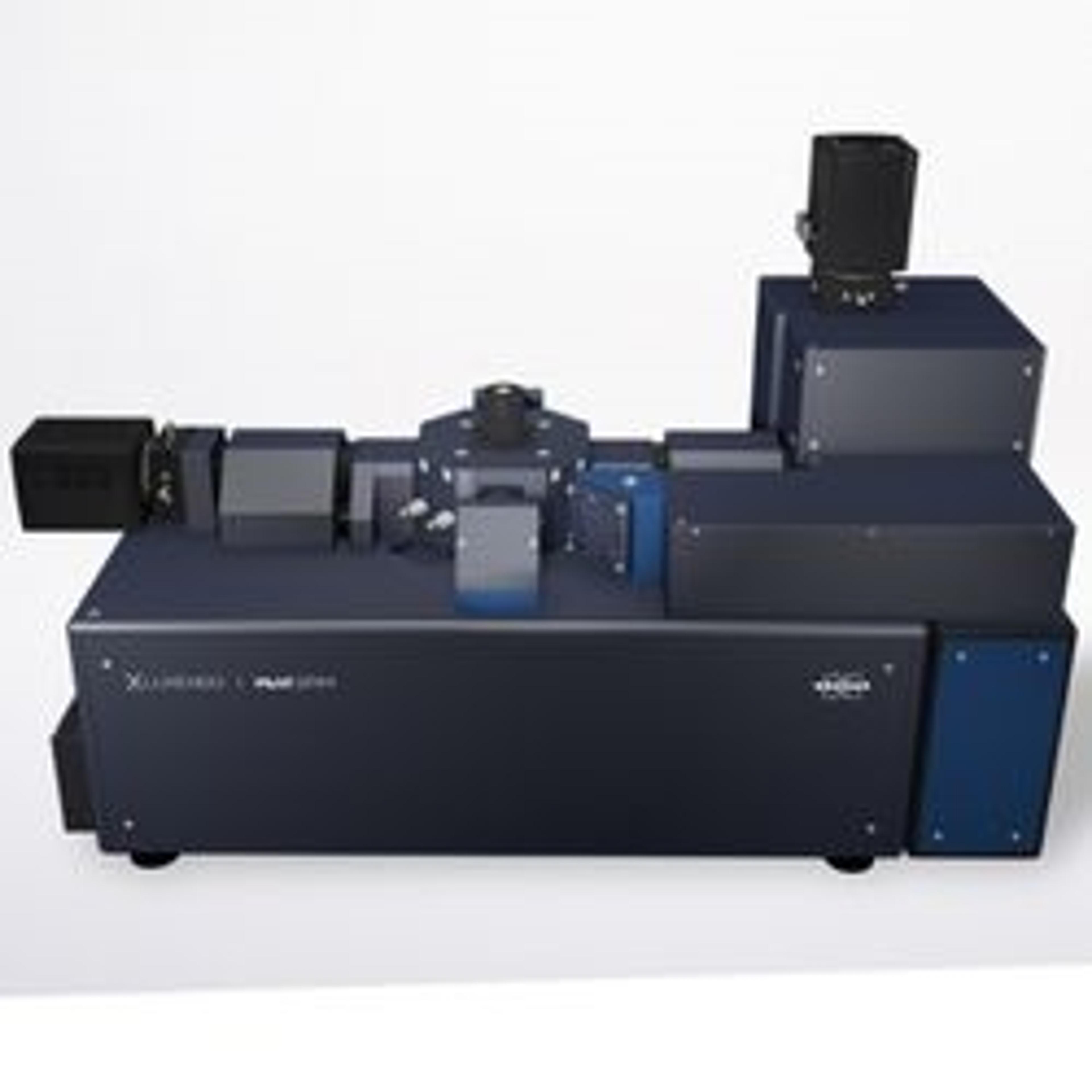
MuVI SPIM
Bruker Fluorescence MicroscopyBruker's MuVi Selective-Plane Illumination Microscope (SPIM) is the fastest multi-angle view system on the market. MuVi incorporates years of Luxendo light-sheet technology innovations to provide high-speed volumetric acquisition for analyzing dynamic processes in delicate live specimens, as well as offering compatibility with any clearing method. A variety of other advanced hardware and software integrations can be easily configured to support evolving multidimensional experimental needs.