Comparison of the effects of pharmaceutical compounds on tumor cells in 2D and 3D in vitro models using label-free, quantitative 4 dimensional holographic imaging

28 Aug 2019

Development of in vitro models for the evaluation of drugs represents a useful approach as in vivo studies may be costly and time consuming. Ideal models should take into account the effects of the cellular microenvironment, which includes the extra-cellular matrix, stroma and neighboring cells. In this poster, the HoloMonitor® M4 from Phase Holographic Imaging was used for label-free long-term kinetic cellular analysis, in order to compare the effects of pharmaceutical compounds on tumor cells.

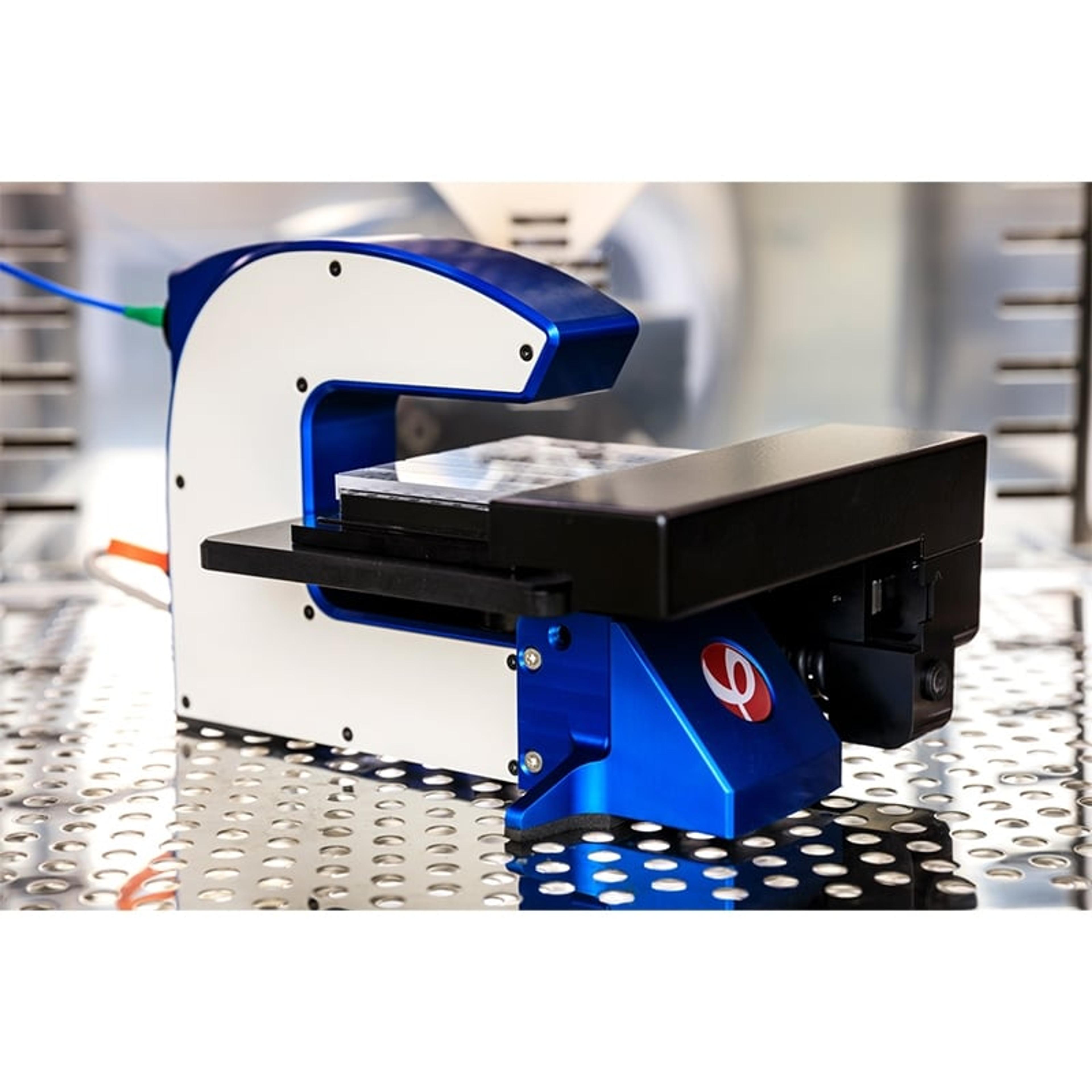
HoloMonitor® M4 Live Cell Analysis System

Phase Holographic Imaging (PHI)

The HoloMonitor ® live cell imaging system enables long-term non-invasive analysis of cell cultures within your standard incubator. Cell biologists worldwide use PHI’s label-free cell imager to automatically obtain accurate live-cell morphology, migration, and proliferation data as well as multi-well time-lapse videos, down to a single-cell level in real-time.

(19)

Links

Tags

Cell / Tissue CultureCell culture or tissue culture is used to study the biology of cells or tissues and to isolate cellular products in an environment which can be manipulated and well defined. Accurately control your culture environment with bioreactors or culture incubators, bind your cells to a surface or together with an extracellular matrix. Distinguish cell types with differential media or proliferate cells with certain characteristics using selective media. Enrich your media with supplements such as growth factors, sera and vitamins. Find the best cell and tissue culture products, kits and equipment in our peer-reviewed product directory: compare products, check customer reviews and receive pricing direct from manufacturers.CytometryCytometry is a broad term for the quantitative analysis of cells and cell systems. The most popular methods to study cytometry are flow cytometry and image cytometry.Cell ImagingCell imaging can be achieved using a number of techniques including confocal microscopy, transmission electron microscopy, atomic force microscopy, and light sheet microscopy.Cell AnalysisThe analysis of cells allows researchers to understand the factors which contribute to cell health and function. These influencing processes can then be predicted and altered, leading to the development of medication and disease treatments.Drug TestingIt is essential to test the efficacy and toxicity of drugs at every stage of their development to ensure that a pharmaceutical product is fit for purpose and safe for use before reaching the stage of mass production and distribution.ImagingImaging techniques are essential for obtaining visual representations of samples to understand structures, processes, and function in biological, chemical, and physical research. These tools range from traditional light microscopy to advanced imaging modalities like MRI and electron microscopy, providing researchers with valuable data for diagnostics, drug discovery, and material analysis. Explore imaging solutions in our peer-reviewed product directory to compare products, check reviews, and get pricing directly from manufacturers.3D Imaging3D imaging technologies allow for the visualization and analysis of three-dimensional structures at high resolution. These systems are used in fields like molecular biology, material science, and medical diagnostics. 3D imaging can be applied to visualize cells, tissues, and organs, providing valuable insights into their structure and function. Browse our peer-reviewed product directory to find the best 3D imaging solutions, compare products, check reviews, and get pricing directly from manufacturers.PharmaceuticalsPharmaceuticals are medicinal drugs used in healthcare to diagnose, prevent, cure and treat illnesses. Pharmaceuticals that are excreted after use appear in wastewater and can have detrimental effects on the environment.Label-Free Analysis