Characterization of Estrogen Receptor α Phosphorylation Sites in Breast Cancer Tissue Using the SNAP i.d® 2.0 System

29 Mar 2017

A major focus of breast cancer research is to understand the mechanisms responsible for disease progression and drug resistance. Toward that end, it has been found that approximately two thirds of all human breast carcinomas overexpress the Estrogen Receptor α (ERα) protein and it remains the primary pharmacological target for endocrine therapy. In this application note, the SNAP i.d.® 2.0 Protein Detection System for multi-slide screening with immunohistochemistry (IHC) is utilized to perform screening of an extensive panel of different breast cancer patient samples and other non-breast cancer tissue microarray (TMA) slide samples, so as to determine their relevance to disease.

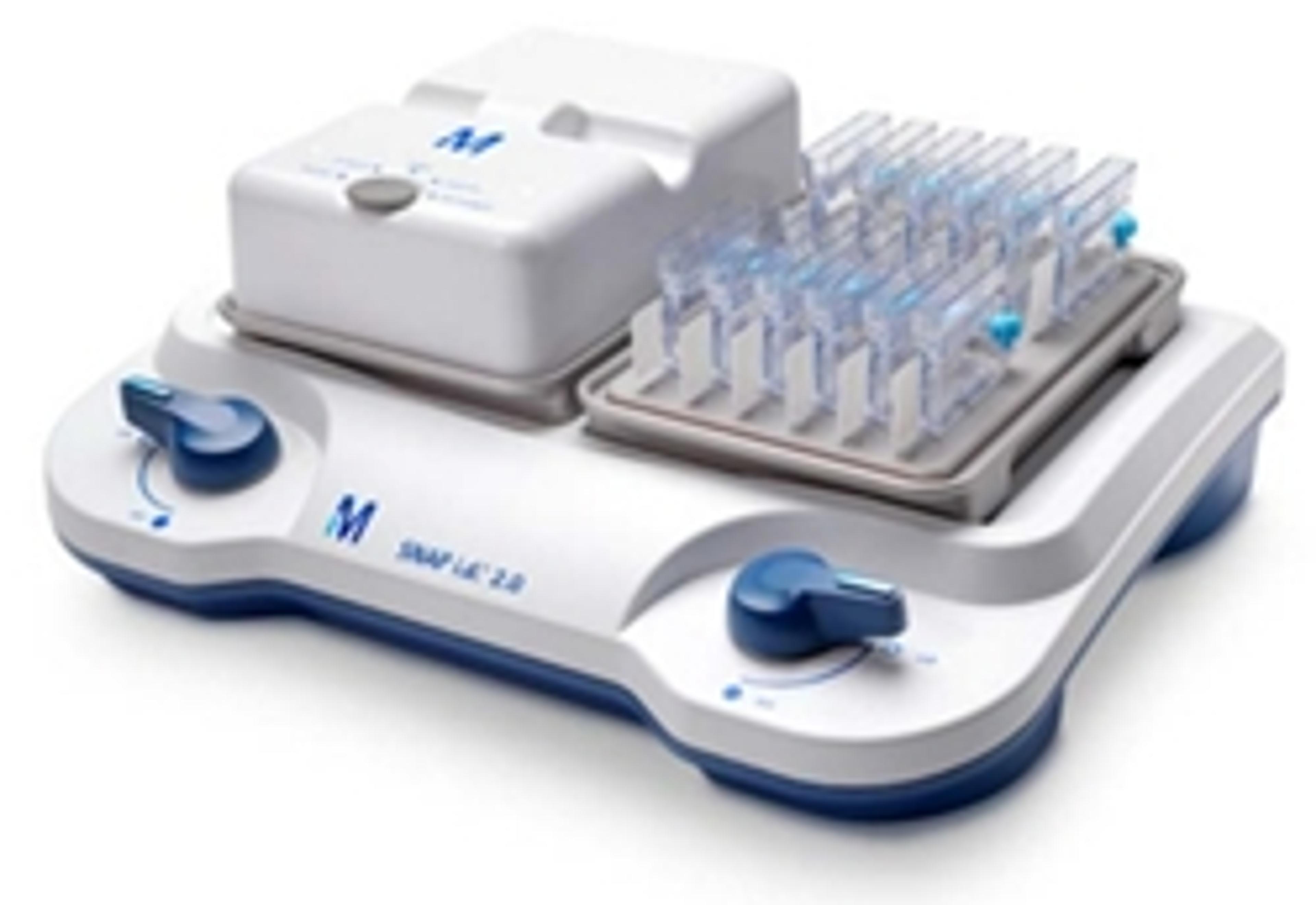
SNAP i.d. 2.0 IHC System

Merck

Issues with Your Tissues? Process all Your Slides in a SNAP for Streamlined IHC. The SNAP i.d.® 2.0 Protein Detection System for Immunohistochemistry (IHC) introduces a new capability to the innovative, vacuum-driven SNAP i.d.® 2.0 system. The IHC slide holders allow you to block, probe, and stain up to 12 tissue slides per side (24 slides if both sides are used). Reduced handling time and multiple-slide processing make this system ideal for when you are optimizing antibody conditions and protocols.With two individually controlled sides, the system base allows for independent, vacuum-driven processing of either one or two IHC frames. Each of the IHC frames can process between 1 to 12 glass slides through independent vacuum ports.Each slide holder has an injection/recovery port that enables the manual addition, as well as the removal and recovery, of small volumes of antibodies or reagents; reagents can also be flushed using the vacuum feature if conservation is not a priority.Benefits: Eliminates the need for pap pens Antibodies can be collected and reused Slide handling time is significantly decreased Less time spent on wash steps Parallel processing of multiple slides Features: Flexibility of multiple slide configurations enables the processing of 1 to 24 slides at a time Compatible with standard IHC slides and protocols Compatible with diverse tissue preparations including formalin-fixed or fresh frozen samples Intuitive format Incorporates blocking, washing, and antibody incubation and labelling steps Systematizes handling multiple slides without the cost of automation Test tracker feature on frame cover helps keep track of IHC steps

(1)

Links

Tags

AntibodiesAntibodies are used in techniques such as confocal and fluorescence microscopy, flow cytometry, ELISA, ELISPOT, immunohistochemistry, western blotting and immunopreciptation. Select specific antigen reactivity, high specific affinity, low non-specific binding, monoclonal or polyclonal, primary or secondary antibodies and associated conjugates such as an enzyme or dye for visualization.ProteomicsProteomics is the systemic bioinformatics study of proteins and amino acids, including their structure, size, function and identification. Tools used in proteomics include chromatography, blotting and gels, protein arrays, mass spectrometry and ELISA and associated analysis software. Analyzers and proteomic systems should be sensitive, high resolution, fast and may be automated for high-throughput.ImmunologyImmunological techniques measure and characterize immune responses. Immunology kits and analysis systems often use techniques such as ELISA, radioimmunoassay (RIA) and immunodiffusion assays, Immunohistochemistry, and flow cytometry. Immunologists use equipment such as flow Cytometers, plate readers, plate washers and fluorescent microscopes.Cellular PathologyCellular Pathology deals with the microscopic analysis of tissue samples and cells. Sample preparation and processing includes fixation, staining, sectioning and slide mounting, using equipment such microtomes and cryostats. In choosing immunohistochemistry and immunocytochemistry kits, consider chromogens, staining method, antibodies, microscopes and imaging.ImmunohistochemistryImmunohistochemistry (IHC) is a technique used to detect specific proteins in tissue samples by using antibodies that bind to target antigens. IHC is widely applied in pathology, immunology, hematology and cancer diagnostics. Explore IHC tools in our peer-reviewed product directory; compare products, check reviews, and get pricing directly from manufacturers.Cancer DiagnosticsThere are a wide variety of diagnostic tests for cancer available, and this range continues to expand as our knowledge of cancer improves. Current diagnostic methods include biopsy, imaging and blood tests for known biomarkers. New methods in research development include liquid biopsies and cancer breathalyzers.Cancer ResearchCancer research aims to understand the mechanisms of cancer development and progression to improve prevention, diagnosis, and treatment. From molecular biology to clinical trials, research spans a wide range of disciplines, including immunotherapy, targeted therapies, and drug discovery. Explore the best cancer research products in our peer-reviewed product directory; compare products, check reviews, and get pricing directly from manufacturers.Pathogen DetectionPathogen detection is the process of identifying harmful microorganisms, including bacteria, viruses, and fungi, that can cause diseases. Accurate pathogen detection is critical for food safety, public health, and clinical diagnostics. Explore pathogen detection products in our peer-reviewed product directory; compare products, check reviews, and get pricing directly from manufacturers.Breast CancerBreast cancer is a type of cancer that begins in the cells of the breast and can spread to other parts of the body. Early detection, accurate diagnosis, and targeted treatments are essential for improving patient outcomes. Browse our peer-reviewed product directory to find the best breast cancer research and diagnostic tools, compare products, check reviews, and get pricing directly from manufacturers.AACRThe American Association for Cancer Research (AACR) is a leading organization focused on cancer research and treatment. It provides a platform for scientists, healthcare professionals, and industry leaders to share knowledge and collaborate in the fight against cancer.
Characterization of Estrogen Receptor α Phosphorylation Sites in Breast Cancer Tissue Using the SNAP i.d® 2.0 System