ResourceLife Sciences
Automated fluorescence imaging and quantification of cell viability
10 Apr 2025Discover how automated fluorescence imaging with the Thermo Fisher Scientific EVOS M7000 and LIVE/DEAD Cell Imaging Kit, paired with advanced image analysis software, transforms cell viability analysis. This powerful combination enables high-throughput, statistically robust data generation in just minutes, streamlining workflows and enhancing efficiency.
Related products
Request Quote for All Products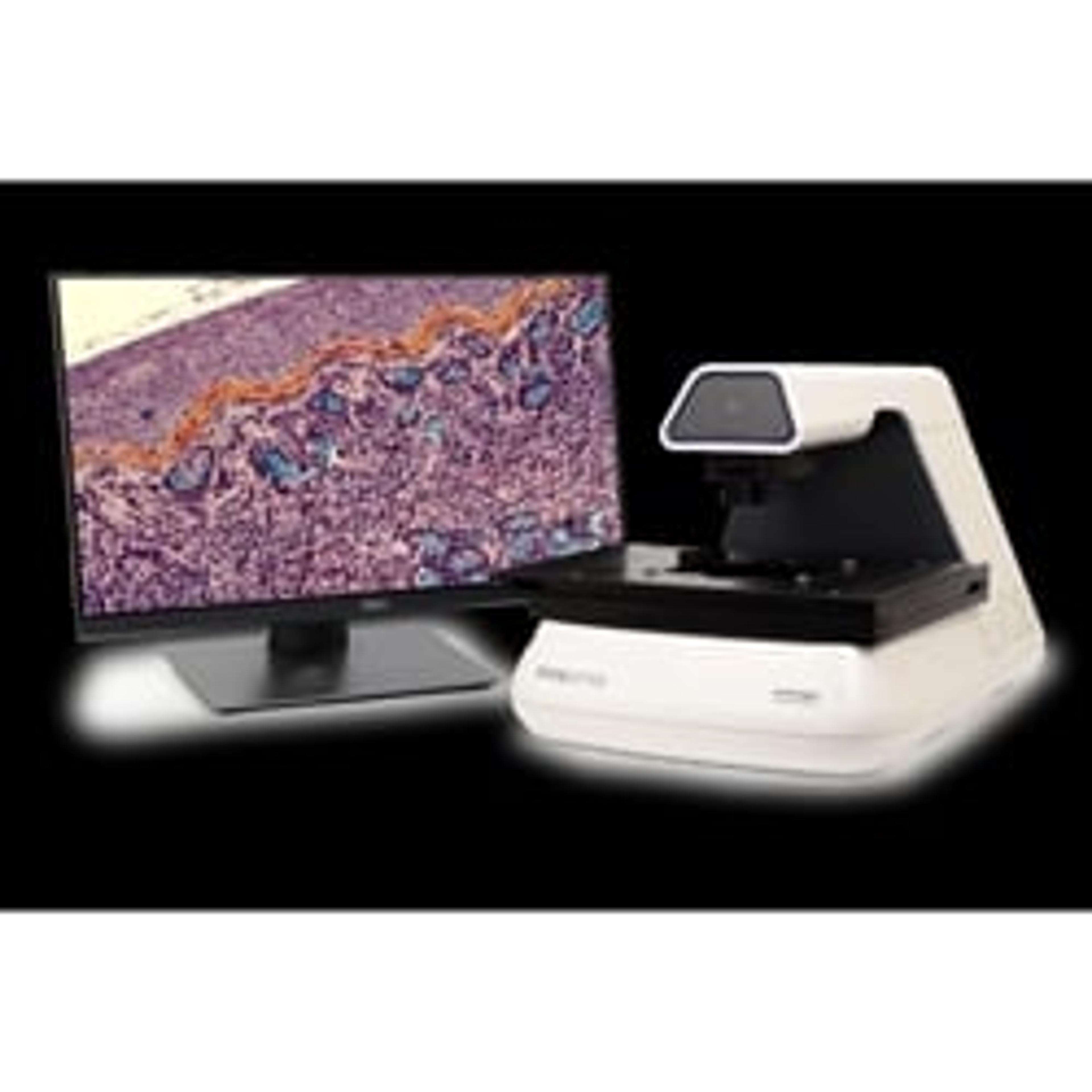
EVOS™ M7000 Imaging System
Thermo Fisher ScientificHigh-performance fully-automated inverted multi-channel fluorescence and transmitted light imaging system.
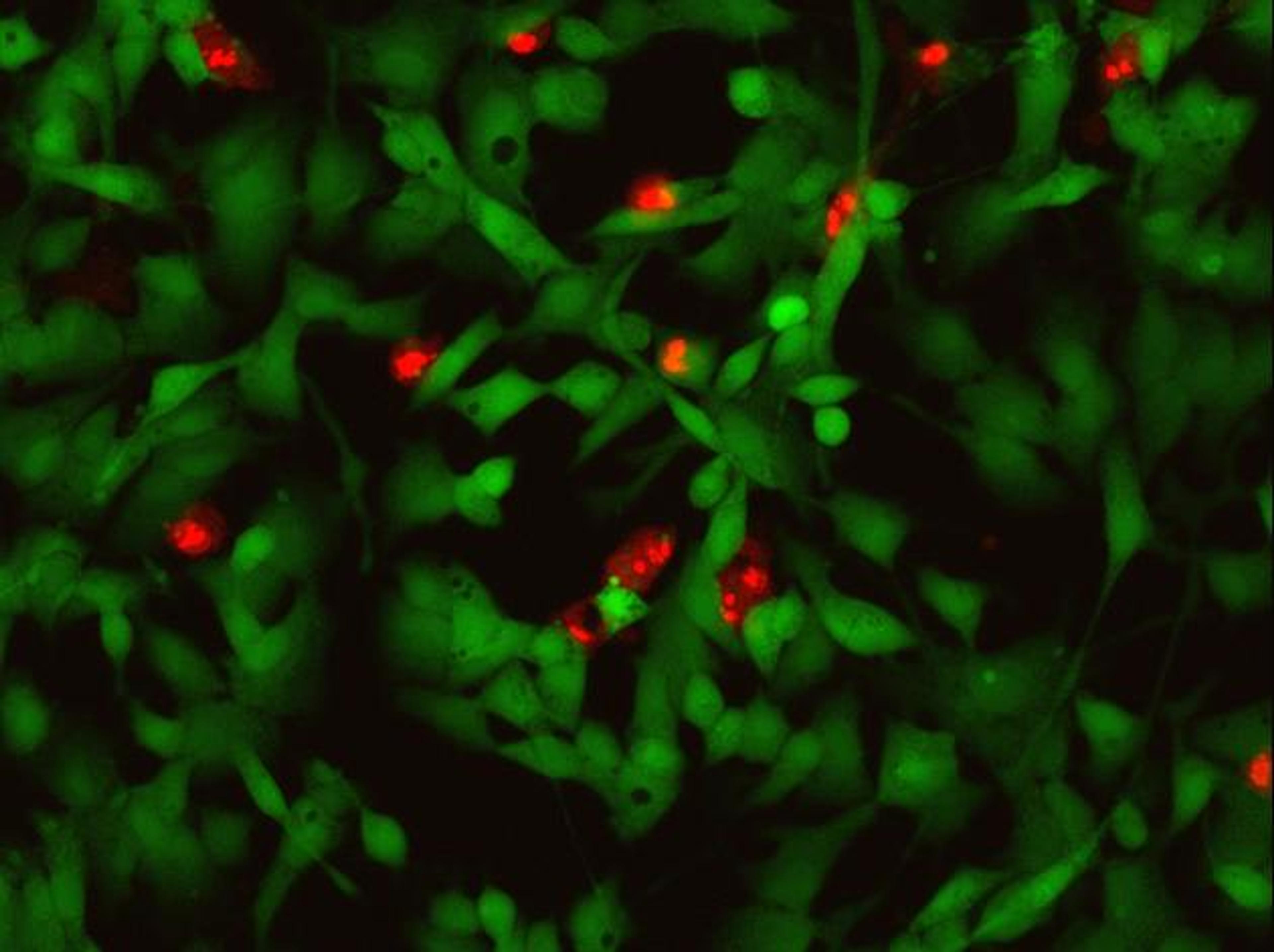
Invitrogen™ LIVE/DEAD™ Cell Imaging Kit (488/570)
Thermo Fisher ScientificSensitive two-color fluorescence cell viability assay optimized for FITC and Texas Red filters

Invitrogen™ Celleste Image Analysis Software
Thermo Fisher ScientificImage processing, manual and automatic measurements over multiple channels, to segmentation and classification tools that help you transform images into quantitative data
Links
Tags
Cellular PathologyCellular Pathology deals with the microscopic analysis of tissue samples and cells. Sample preparation and processing includes fixation, staining, sectioning and slide mounting, using equipment such microtomes and cryostats. In choosing immunohistochemistry and immunocytochemistry kits, consider chromogens, staining method, antibodies, microscopes and imaging.Cell ViabilityCell viability assays assess the health of cells and their ability to proliferate. These tests are crucial in drug development, toxicity testing, and cell-based research. Common methods for assessing viability include MTT, live/dead assays, and flow cytometry. Browse our peer-reviewed product directory to find the best tools for cell viability assays, compare products, check reviews, and get pricing directly from manufacturers.AutomationAutomation in laboratories and manufacturing processes enhances efficiency, precision, and scalability by reducing the need for manual intervention. It plays a critical role in improving productivity, minimizing human error, and accelerating workflows in fields like diagnostics, drug development, and industrial testing. Automation technologies include robotic systems, automated liquid handlers, and process control systems that streamline complex tasks and ensure consistent, reproducible results. Explore our peer-reviewed product directory to discover the best automation solutions, compare options, read user reviews, and get prices directly from manufacturers.Fluorescence Imaging