ResourceLife Sciences
Automated Cell Dispensing and Image-Based Spheroid Formation Tracking
20 Aug 2014This application note demonstrates how dispensing and imaging procedures are automated to facilitate higher throughput in 3D HDP processing, using the MultiFlo™ FX and Cytation™ to dispense cell suspensions into hanging drop plates and track spheroid formation via digital widefield imaging.
Related products
Request Quote for All Products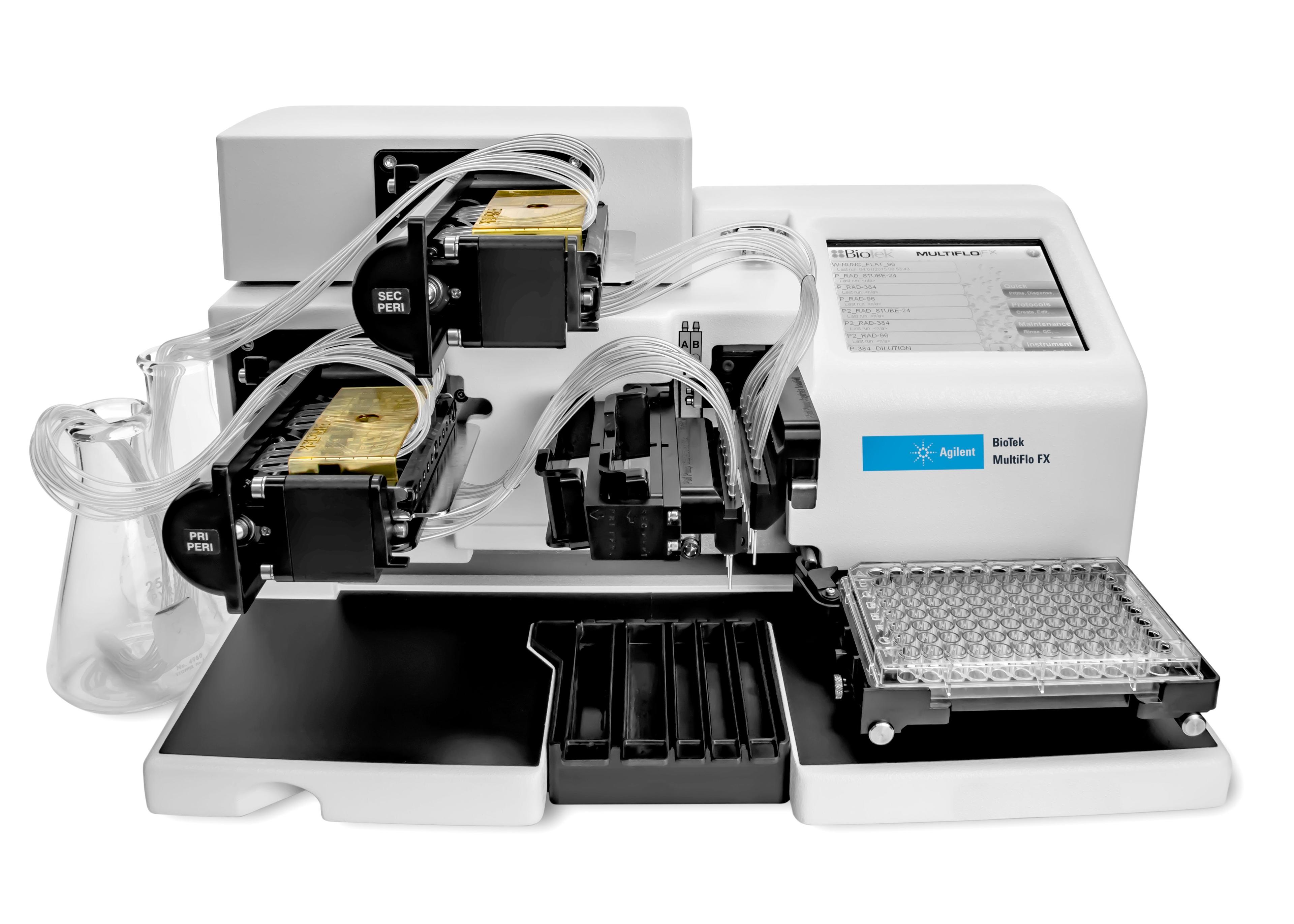
Agilent BioTek MultiFlo FX Multimode Dispenser
Agilent TechnologiesMultiFlo FX can dispense up to 4 independent reagents into 6- to 1536-well microplates.
Links
Tags
In Vivo Imaging Systems<i>In vivo</i> imaging systems, including pre-clinical imaging systems and medical imaging systems are used to non-invasively visualize and capture images of live animals and plants. Monitor the natural processes or diseases of your subjects using small-animal pre-clinical imaging systems, including single photon positron emission tomography (SPECT), positron emission tomography (PET), computed tomography (micro-CT), magnetic resonance imaging (MRI), X-ray radiography, ultrasound, fluorescence and bioluminescence imagers. Multimodal systems and software solutions are also available for correlative analysis of organ, tissue, cell, or molecular-level processes. Find the best in vivo imaging products in our peer-reviewed product directory: compare products, check customer reviews and receive pricing direct from manufacturers.Cell / Tissue CultureCell culture or tissue culture is used to study the biology of cells or tissues and to isolate cellular products in an environment which can be manipulated and well defined. Accurately control your culture environment with bioreactors or culture incubators, bind your cells to a surface or together with an extracellular matrix. Distinguish cell types with differential media or proliferate cells with certain characteristics using selective media. Enrich your media with supplements such as growth factors, sera and vitamins. Find the best cell and tissue culture products, kits and equipment in our peer-reviewed product directory: compare products, check customer reviews and receive pricing direct from manufacturers.SpheroidsSpheroids are clusters of cells that have been grown in 3D culture to be used as <i>in vitro</i> model systems. These 3D microtissues can be used for toxicology testing, DMPK studies and many other applications involving cell analysis.FluorescenceThe emission of fluorescence occurs when a photon of energy is supplied to a fluorescent chemical compound by an external source, causing it to become excited. Fluorescence can be detected and measured for different purposes using microplate readers, fluorescence microscopes, fluorescence scanners, and flow cytometers.