Applications of cryo-electron microscopy

Cryo-electron microscopy (cryo-EM) allows the structural analysis of protein complexes flash-frozen in their near native states. You can now directly visualize not just large macromolecules but also smaller proteins complexes, including membrane proteins. This powerful technique can be used to complement traditional methods, such as X-ray crystallography (XRD) or nuclear magnetic resonance (NMR) for structure based drug discovery

22 Sept 2019

In this application note, Thermo Fisher Scientific introduces the versatility of cryo-EM and the variety of benefits made possible by this method. This application outlines how cryo-EM provides a better insight into all classes of biomolecules, including proteins that are difficult to work with.

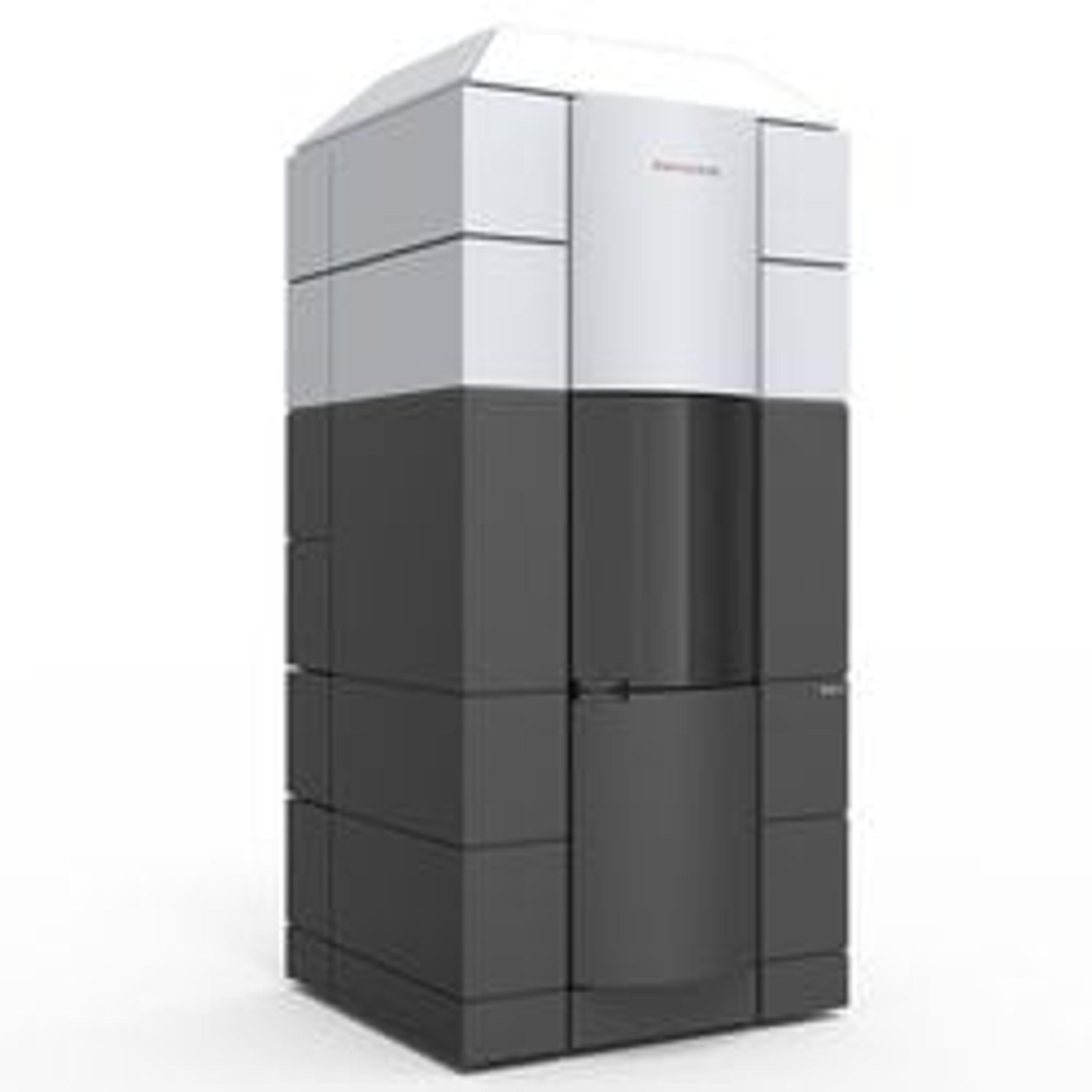
Krios G4 Cryo-TEM for Life Sciences

Thermo Fisher Scientific

The Thermo Scientific Krios G4 Cryo-Transmission Electron Microscope (Cryo-TEM) is a compact TEM. The instrument can be installed in labs with a ceiling height below 3.04 m (~10 ft), as the microscope height it below 3 meters. The Krios G4 Cryo-TEM has improved ergonomics for easier sample exchange. Data acquisition set up is easier and faster thanks to enhanced automation, systematic user guidance and advanced performance monitoring. Simultaneo

(0)

Links

Tags

ProteomicsProteomics is the systemic bioinformatics study of proteins and amino acids, including their structure, size, function and identification. Tools used in proteomics include chromatography, blotting and gels, protein arrays, mass spectrometry and ELISA and associated analysis software. Analyzers and proteomic systems should be sensitive, high resolution, fast and may be automated for high-throughput.Electron MicroscopyElectron microscopes (EM) are used to create high-resolution images of samples at the nanoscale by means of an accelerated beam of electrons as a source of illumination. Types of electron microscope include scanning electron microscopes (SEM), transmission electron microscopes (TEM), scanning transmission electron microscopes (STEM) and cryo-electron microscopes. Focused ion beam (FIB) microscopes are useful for modifying or milling a sample surface with nanometer precision, as well as imaging. Find the best electron microscopes in our peer-reviewed product directory: compare products, check customer reviews and receive pricing direct from manufacturers.MicroscopyMicroscopy is a technique used to observe small objects in detail, from cells to materials, using light or electron microscopes. It enables researchers to examine structures with high resolution, aiding in fields such as biology, medicine, and materials science. With advanced microscopy techniques, scientists can gain insights into cellular processes, tissue structures, and material properties. Explore the best microscopy solutions in our peer-reviewed product directory, compare products, read customer reviews, and get pricing directly from manufacturers.Drug DiscoveryDrug discovery is the process of identifying potential new medications, involving stages such as target identification, compound screening, and preclinical development. It relies on cutting-edge technologies like high-throughput screening, artificial intelligence, and molecular modeling to accelerate the identification of drug candidates. Drug discovery plays a pivotal role in developing new therapies for diseases ranging from cancer to rare genetic disorders. Browse our peer-reviewed product directory to find the latest drug discovery technologies, compare options, check customer feedback, and get pricing directly from manufacturers.Drug DevelopmentDrug development refers to the process of bringing a new drug to market.electron microscopy