Alternatives to DAPI Staining: Imaging and Counting Live Cells
Alternatives to DAPI Staining: Imaging and Counting Live Cells
16 Nov 2015DAPI (4’, 6-diamidino-2-phenylindole) is a fluorescent dye often used to stain nuclear DNA. It is employed in imaging experiments such as fluorescent microscopy, chromosome spreads, FACS, and cell-based assays. However, excitation with UV results in a photoconversion of DAPI that leads to detection of DAPI fluorescence in the FITC/GFP channel of an imaging system, causing errors in interpretation of results. DAPI also requires cells to be fixed for maximal staining. In this application note, some alternatives to DAPI staining which require no staining at all and can be used with live cells are shown.
Related products
Request Quote for All Products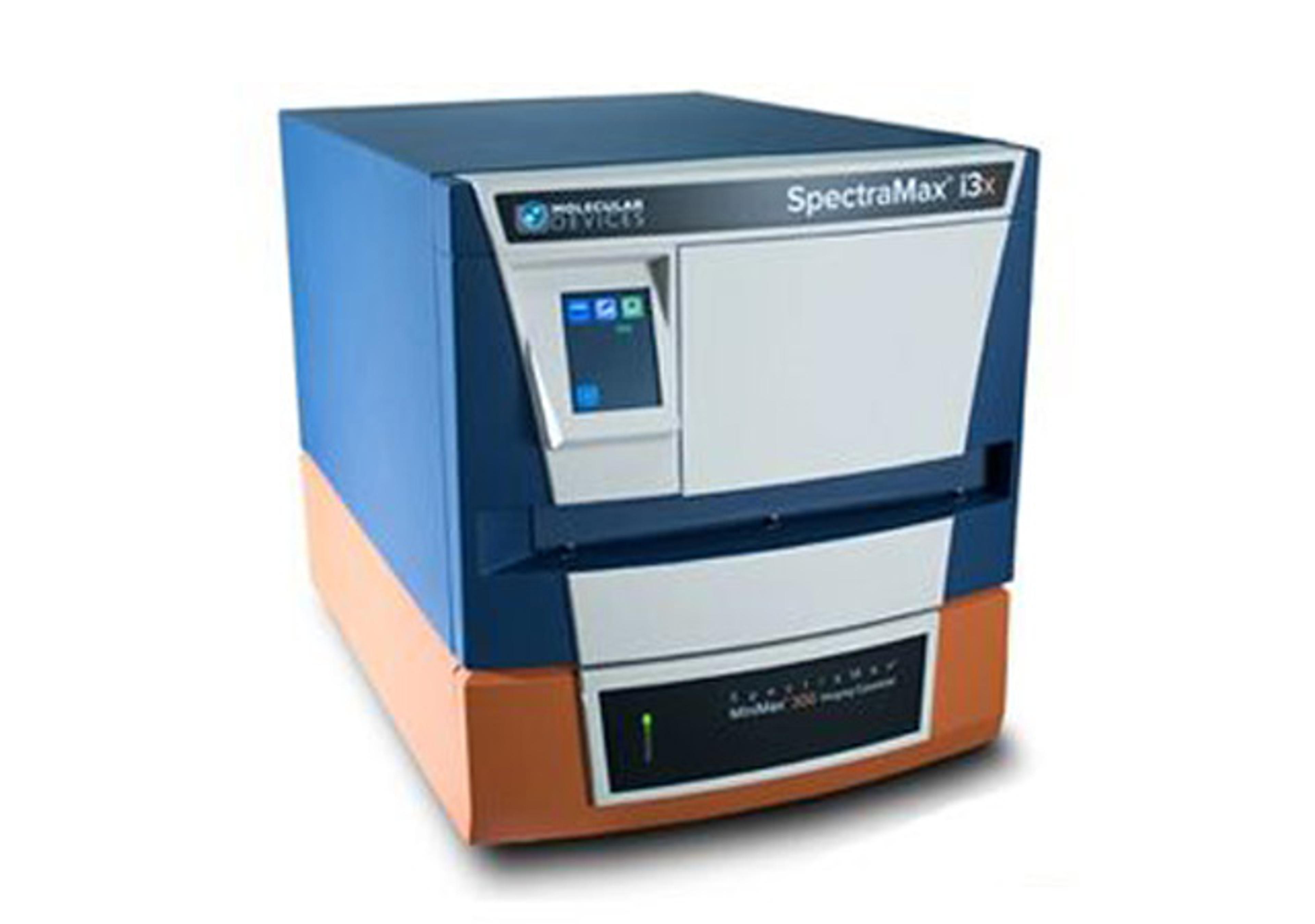
SpectraMax i3x Multi-Mode Microplate Reader with MiniMax Imaging Cytometer
Molecular Devices®Measures Absorbance, Fluorescence, and Luminescence with the added functionality of modular upgrades