Airyscanning: A Novel Approach to Confocal Imaging
21 Nov 2014Confocal laser scanning microscopy is the recognized standard for 3D fluorescence microscopy. This application note describes how Airyscanning releases the full potential of a confocal microscope, achieving resolutions comparable to an extremely small pinhole but with a much better signal-to-noise ratio. It combines excellent optical sectioning performance with flexible scanning strategies for imaging and photomanipulation, making it the method of choice for a vast range of applications.
Related products
Request Quote for All Products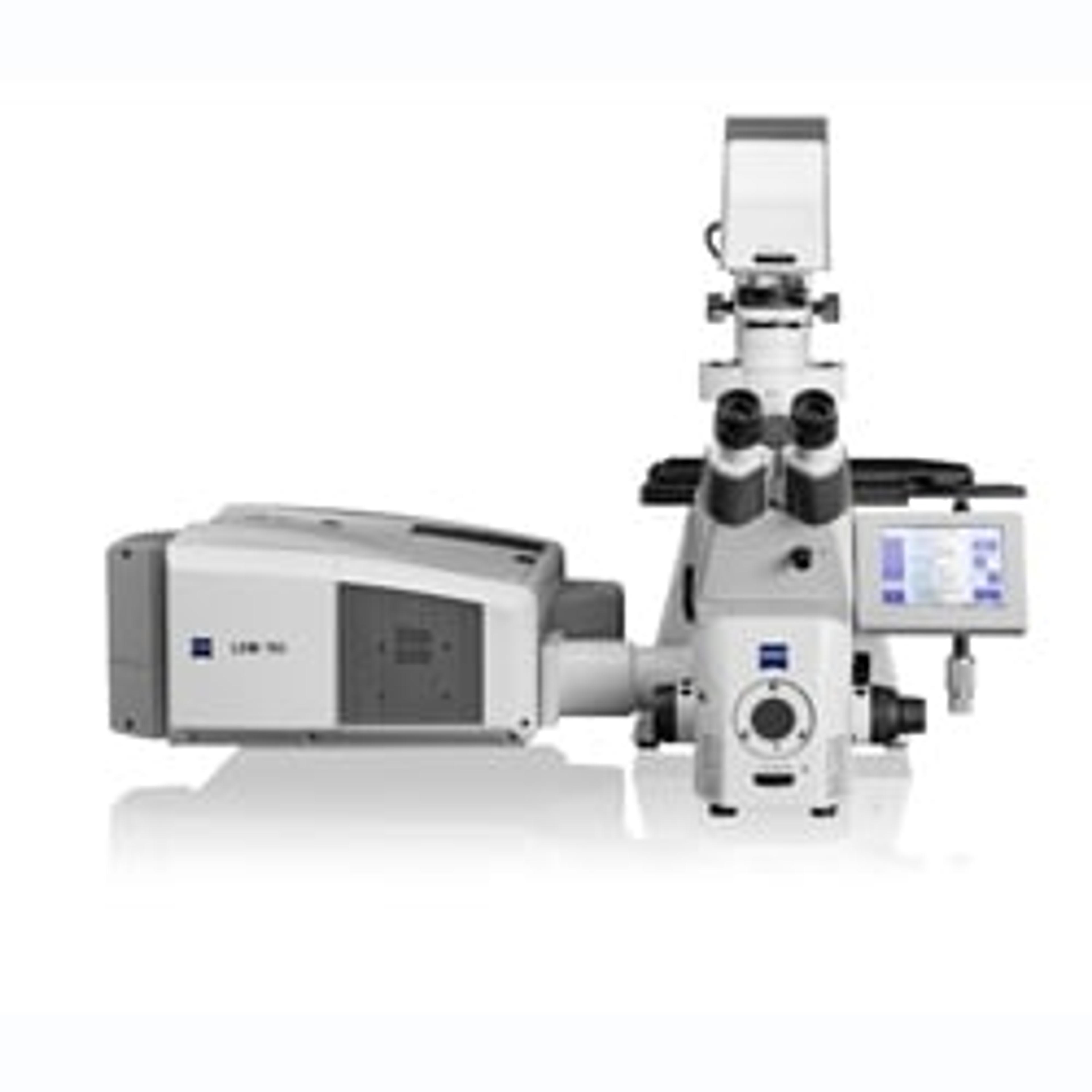
ZEISS LSM 780 Laser Scanning Microscope
ZEISS Research Microscopy SolutionsLSM 780 - Obtain Quantitative Information About Your Cells and Individual Molecules. The sensitivity of LSM 780 is quite simply outstanding. The GaAsP detector achieves 45 percent quantum efficiency compared to 25 percent typically by conventional PMT detectors. This results in accurate details and contrast-rich images of the challenging specimens you encounter in your live cell imaging.The system’s illumination and detection design allows you to simultaneously acquire up to ten dyes. You excite any common fluorophore with up to eight different lasers, detecting the signals with the 32 channel GaAsP detector. LSM 780 is so sensitive, the system even allows photon counting.The maximum possible acquisition speed available to you is determined by the sensitivity of your detection system. When you choose LSM 780, you benefit from new GaAsP detectors that bring a significant improvement on signal-to-noise ratio, allowing you to acquire your image sequences at least twice as fast.Parallel spectral detection also reads 34 channels simultaneously in Lambda mode: you acquire and separate up to ten colors simultaneously.LSM 780 allows you to achieve images of unparalleled detail, even from the most challenging specimens. The superior quality and sensitivity has been recognized by a distinguished R&D 100 Award in 2011.LSM 780 Laser Scanning Microscope Features: 32-channel GaAsP detector with a typical quantum efficiency of 45% – a significant improvement on the 25% typically achieved by classic PMT detectors. Potential for photon counting on GaAsP and the new side PMT. With lasers from 355 nm true UV to 660 nm far red, any dye will be excited optimally. Simple adaptation to changing acquisition strategies via the ZEN software’s Smart Setup. Improved ZEN software with modules for e.g. correlative microscopy, deconvolution and time lapse. Improved dynamics for better visualization of weak signals. Active cooling together with oversampling and photon counting modes deliver improved signal-to-noise ratio.

ZEISS LSM 880 with Airyscan
ZEISS Research Microscopy SolutionsYour New Standard for Fast and Gentle Confocal Imaging