ProductsLife Sciences
STA®-Liatest® D-Di Plus
Diagnostica StagoLess interferences for even more confidence

Less interferences for even more confidence
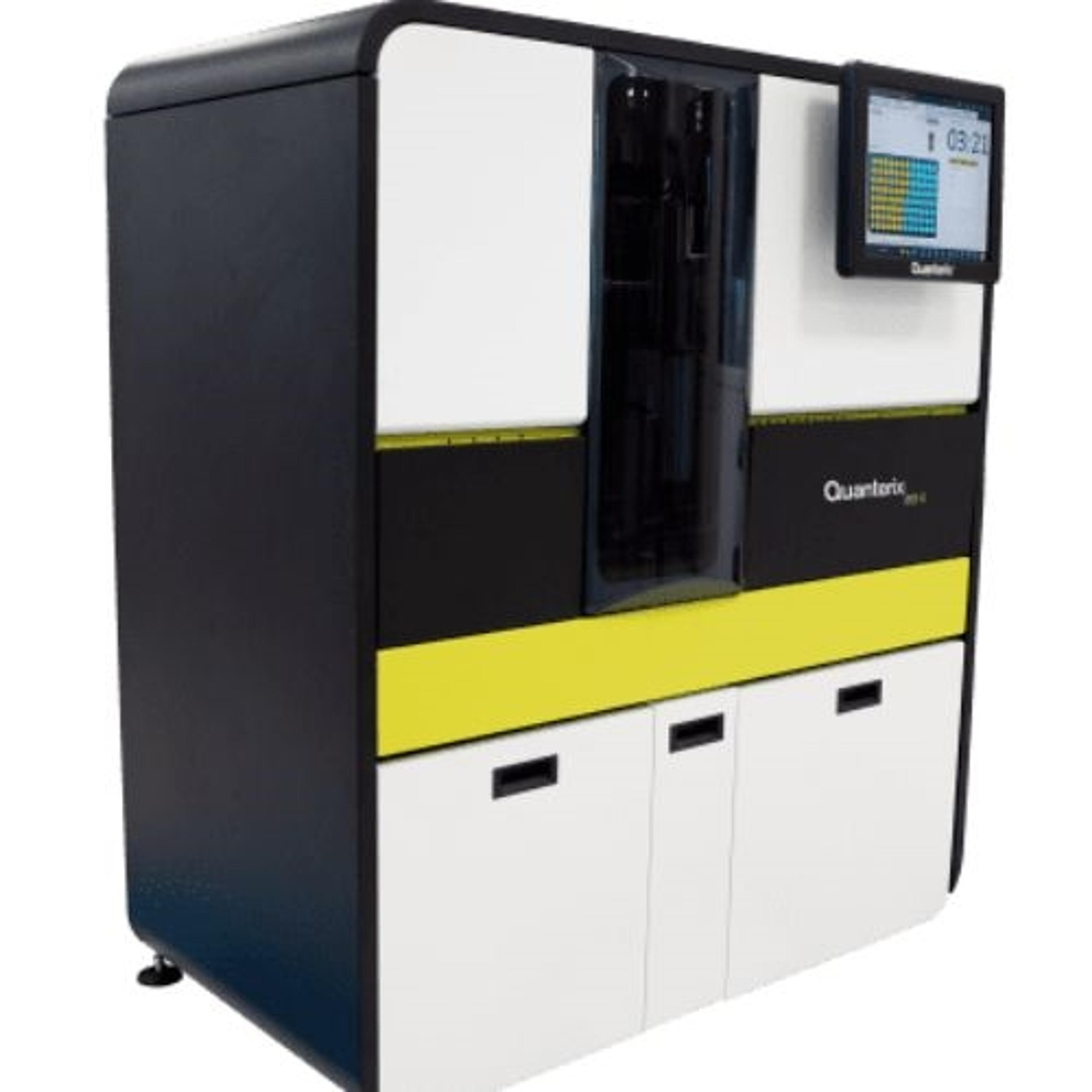
MSD's most affordable imager, with special pricing for academic customers
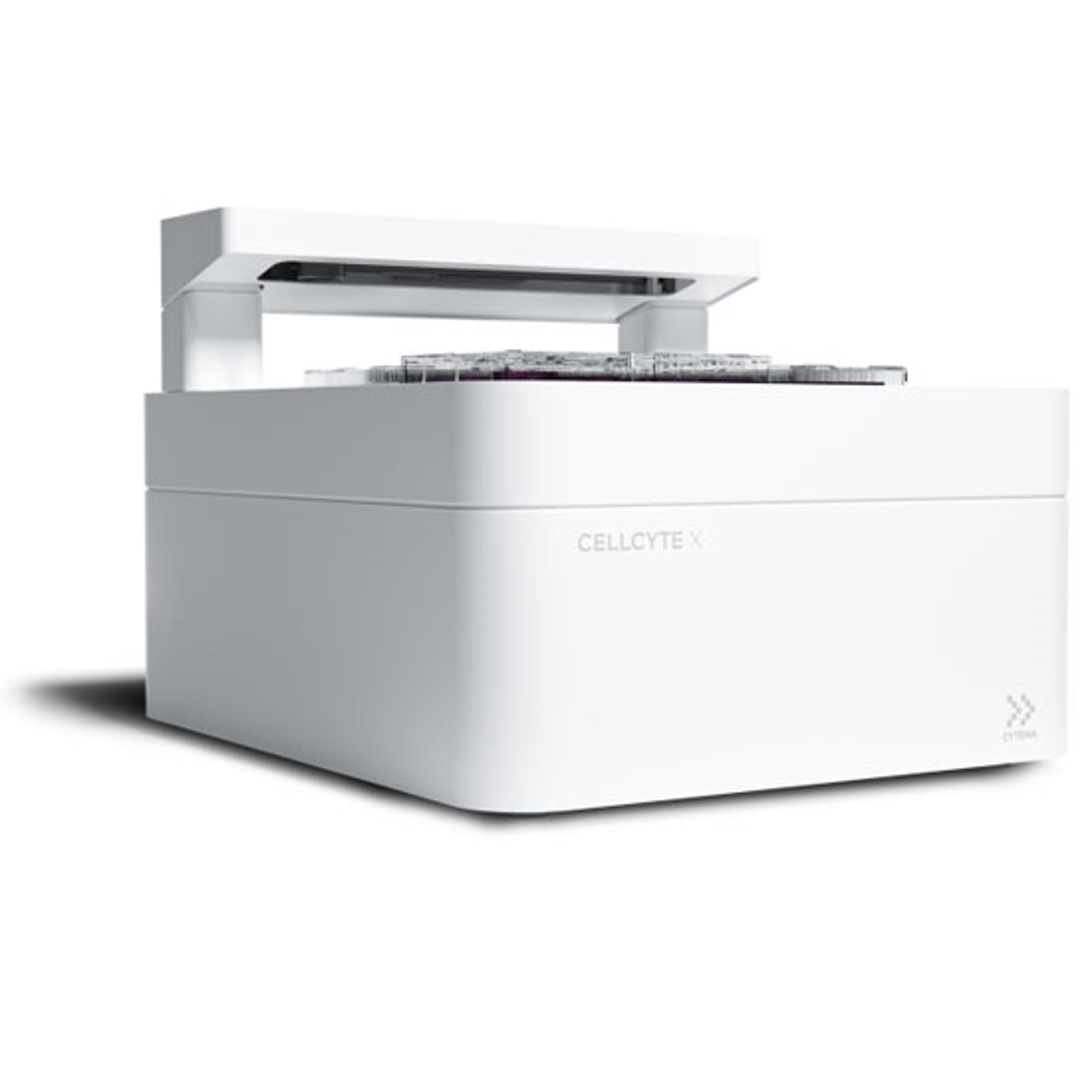
Get the full picture of cell behavior over time.
Sigma′s FLAG Immunoprecipitation kit allows a rapid and efficient immunoprecipitation and elution of an active FLAG-tagged protein.
The RarePlex Panel Kit product line includes immunofluorescence Panel Kits and Developer Kits for circulating tumor cell (CTC) characterization, including enumeration, and specific kits include additional biomarkers for in-depth phenotypic characterization. RarePlex Panel Kits are highly validated circulating tumor cell assays designed to work with the AccuCyte® Sample Preparation System, automated slide staining systems, a…
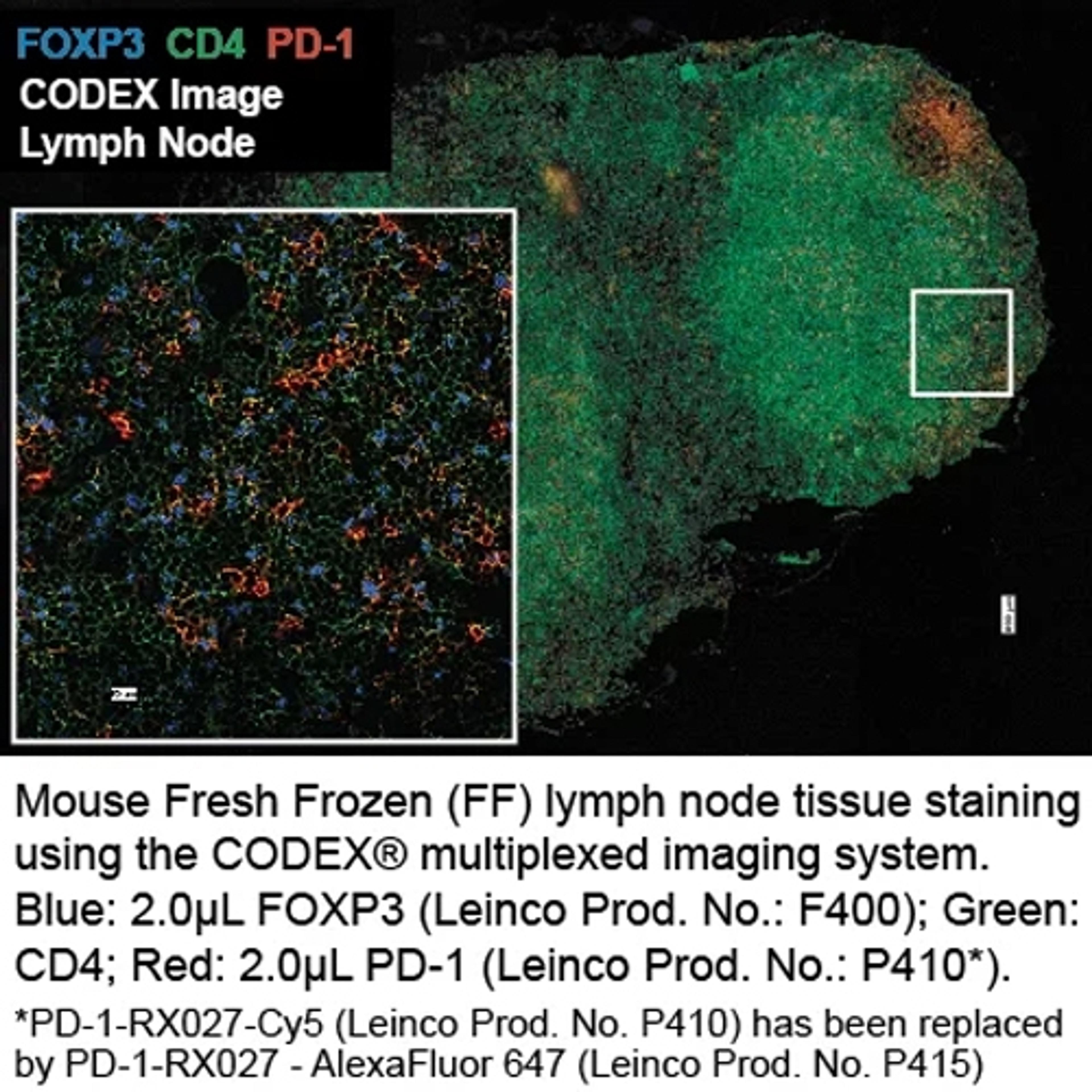
Anti-Mouse PD-1-BX027 (RMP1-30); AlexaFluor 647-RX027
Enzyme Immunoassay for the quantitative determination of PEG-GCSF in serum, plasma and cell culture supernatant.
CytoSections is a verified, reproducible, and renewable positive control source where the target protein's expression is confirmed by western blot and IHC.
Aura® PTx gets you to your best formulation faster! Backgrounded Imaging (BMI) technology to give you accurate count, size, and morphology information about particles in your sample. Couple that with Fluorescence Membrane Microscopy (FMM), which gives you definitive ID of protein aggregates and an easy way to monitor polysorbate degradation.
Our unique combination of custom antibody development, lateral flow development and manufacturing services, signal enhancement technology and our mobile diagnostic platform are shaping the future of lateral flow. Our partnership approach allows you to work with one company to reach your assay goals.
Monoclonal Anti-Claudin-18.2 Antibody, Mouse IgG1 (3B10) was developed to be exquisitely CLDN18.2 specific, characterized as not cross-reacting with CLDN18.1 and without background staining.
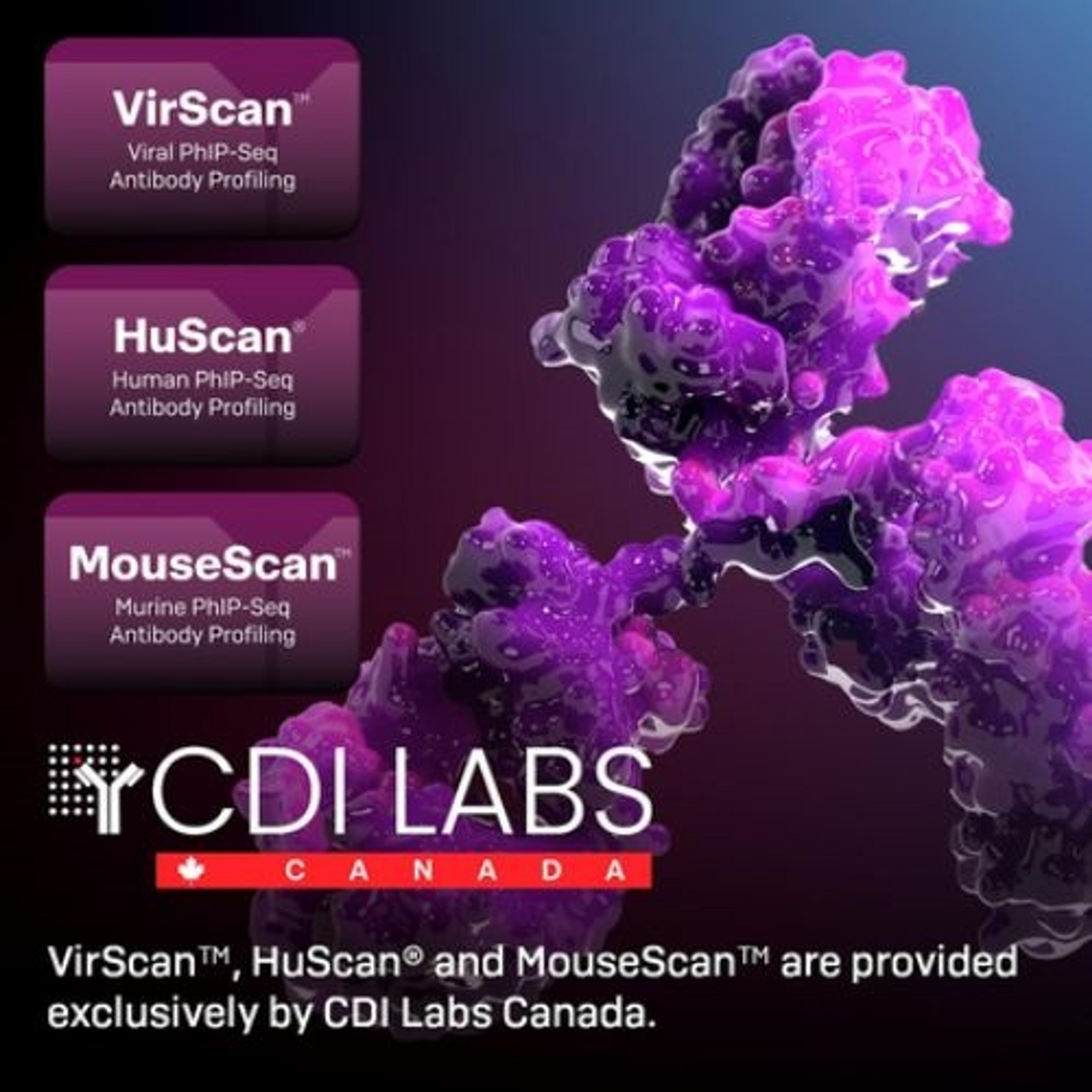
VirScan™, HuScan ® , and MouseScan™ are applications of bacteriophage-display immunoprecipitation sequencing (PhIP-Seq), a powerful method of multiplexed analysis that combines high-throughput DNA sequencing with next-generation proteomics. PhIP-Seq is the highest-coverage antigen-specific assay yet developed, able to detect antibodies versus virtually any antigen via overlapping peptides.
IP-One ELISA assay kit from Cisbio Bioassays - Simple and highly accurate measurement of IP1 IP1, a downstream metabolite of IP3, accumulates in cells following activation of Phospholipase C (PLC) coupled receptors. Among them, the Gq coupled GPCRs represent the most important family of receptors which can activate the β subtype of the PLC family. Other receptor types, like protein tyrosine kinase receptors, antigen or immuno…
Biotin-conjugated anticoagulant for the detection of phosphatidylserine in the outer leaflet of apoptotic cellsProduct Description Detection: Annexin-V-Biotin can be used in immunohistochemistry with a secondary detection system (e.g., Streptavidin-POD conjugate). Sample material: Cell lines and freshly isolated cells Specificity: Annexin-V binds in a calcium-dependent manner to negatively charged phospholipid surfaces, and…
Roche Cell Death Detection ELISA - Photometric enzyme immunoassay for the quantitative in vitro determination of cytoplasmic histone-associated DNA fragments (mono- and oligonucleosomes) after induced cell deathBenefits of Roche Cell Death ELISA: High sensitivity: (5 x 10² cells/ml) Fast performance: (5 - 6 hours) No prelabeling of cells necessary Nonradioactive assay system No species restrictions Easy handling…
Kit for the detection and quantification of apoptotic cell death on a single-cell level by light microscopy in immunocyto- and immunohistochemistry Benefits Sensitive: The maximum intensity of labeling (cell staining) of apoptotic cells is higher than the nick translation method. Fast: The use of fluorescein-dUTP allows analysis of the samples directly after the TUNEL reaction, but before the addition of the secondary d…
Kit for the detection and quantification of apoptotic cell death on a single-cell level by flow cytometry and fluorescence microscopy, and for double labeling with fluorescein-labeled cell markers (TMR red)Benefits Convenient: No secondary detection system required. Accurate: Identification of apoptosis at a molecular level (DNA-strand breaks) and identification of cells at the very early stages of apoptosis. Flexible:…
Application The Cellular DNA Fragmentation ELISA may be applied to measure: Apoptosis by detection of BrdU-labeled DNA fragments in the cytoplasm of affected cells (= nonradioactive alternative to the [3H]-thymidine-based DNA-fragmentation assay). Cell-mediated cytotoxicity by detection of BrdU-labeled DNA fragments released from damaged target cells into the culture supernatant (= nonradioactive alternative to the [3H]-thymid…
Application The kit is used for the detection of BrdU incorporated into cellular DNA by immunocyto/histochemistry. Benefits Safe: No radioisotopes are used. Easy to perform: Follows a standard immunohistochemistry protocol. Sensitive: Denaturation of DNA with nucleases allows for highly sensitive detection of BrdU. Flexible: Allows double-labeling protocols.
Application The kit is used for the detection of BrdU incorporated into cellular DNA using immunofluorescence microscopy. It is used for the detection of DNA synthesis by either in vitro labeling of cells or organ cultures, or by in vivo labeling, in which frozen or paraffin-embedded tissue sections must be prepared prior to fixation. Benefits Safe: No radioisotopes are used. Easy to perform: Follows a standard immunof…
Application The Cell Proliferation ELISA, BrdU (colorimetric) belongs to the second, improved generation of kits for measuring DNA synthesis. It is designed as a precise, fast, and simple colorimetric alternative to quantitate cell proliferation based on the measurement of BrdU incorporation during DNA synthesis in replicating (cycling) cells. Thus, the Cell Proliferation ELISA can be used in many different in vitro cell syst…
ApplicationThe In Situ Cell Proliferation Kit, FLUOS belongs to the second generation of improved kits for the detection of DNA synthesis at the single-cell level (in situ) by immunocyto-/immunohistochemistry. The kit can be used in many different in vitro and in vivo cell systems, for example: Detection and quantification of cells in S-phase Determination of growth fractions for tumors in animal-model systems Detectio…