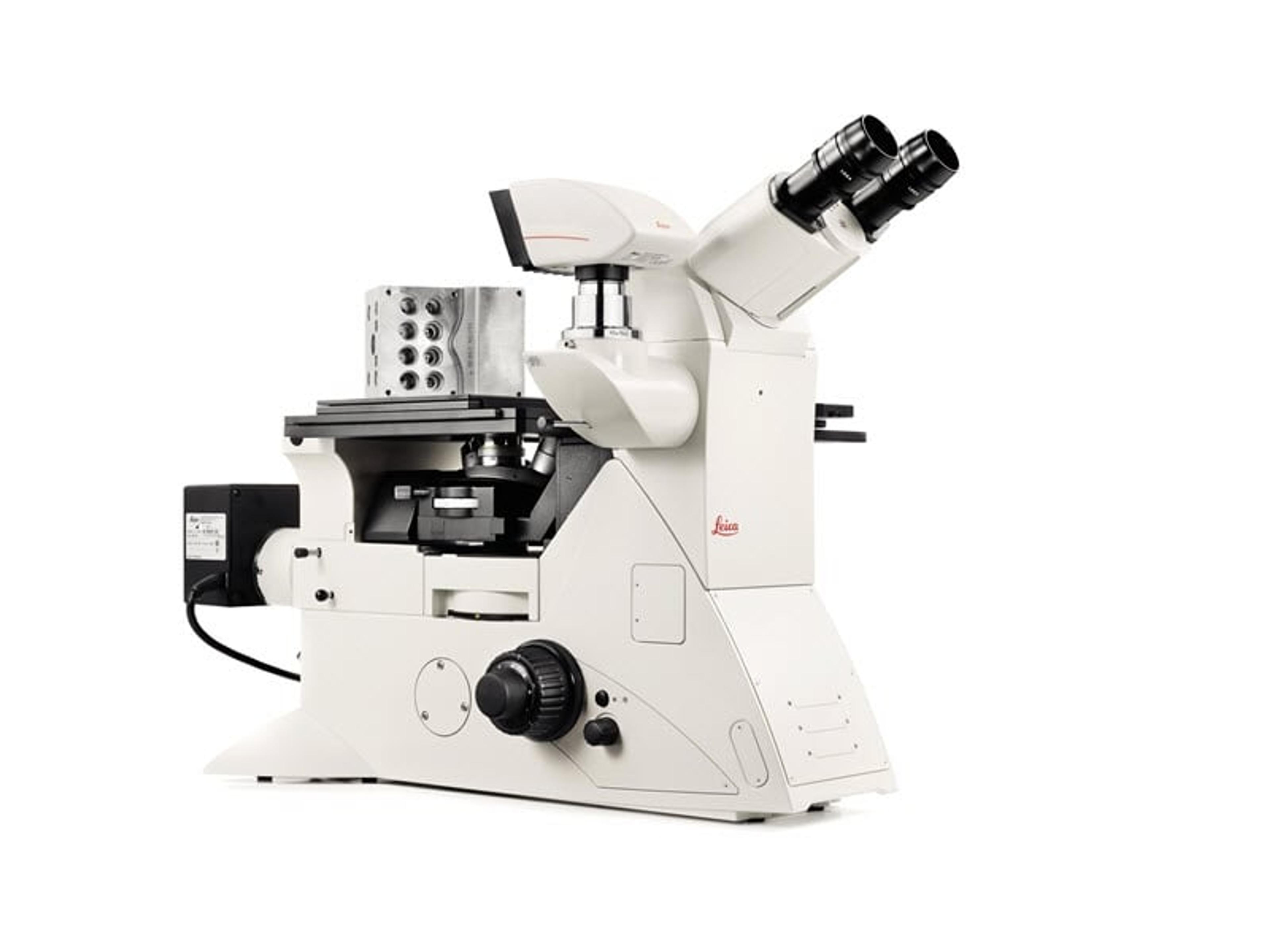
DMi8 M/C/A Inverted Microscopes (Industry)
Leica Microsystems EuropeInverted Microscopes for Industry
Inverted Microscopes for Industry
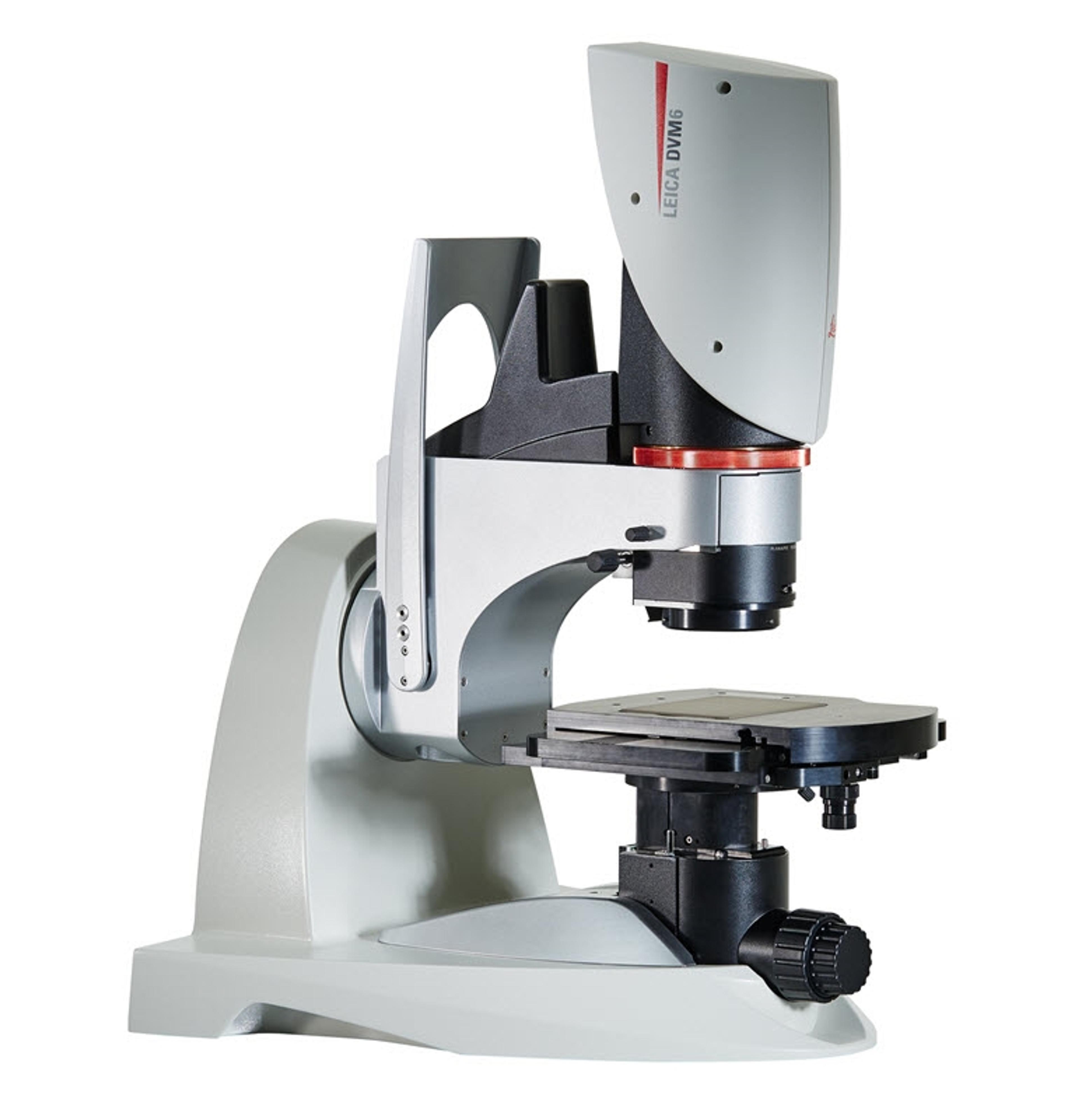
If you work in quality control/assurance, failure analysis, research and development, or in forensics, searching for the detail can take up a lot of your time in microscopy. The DVM6 digital microscope is a fast, reliable and easy to use solution that combines outstanding optics, intuitive operation, and smart software to save you time.
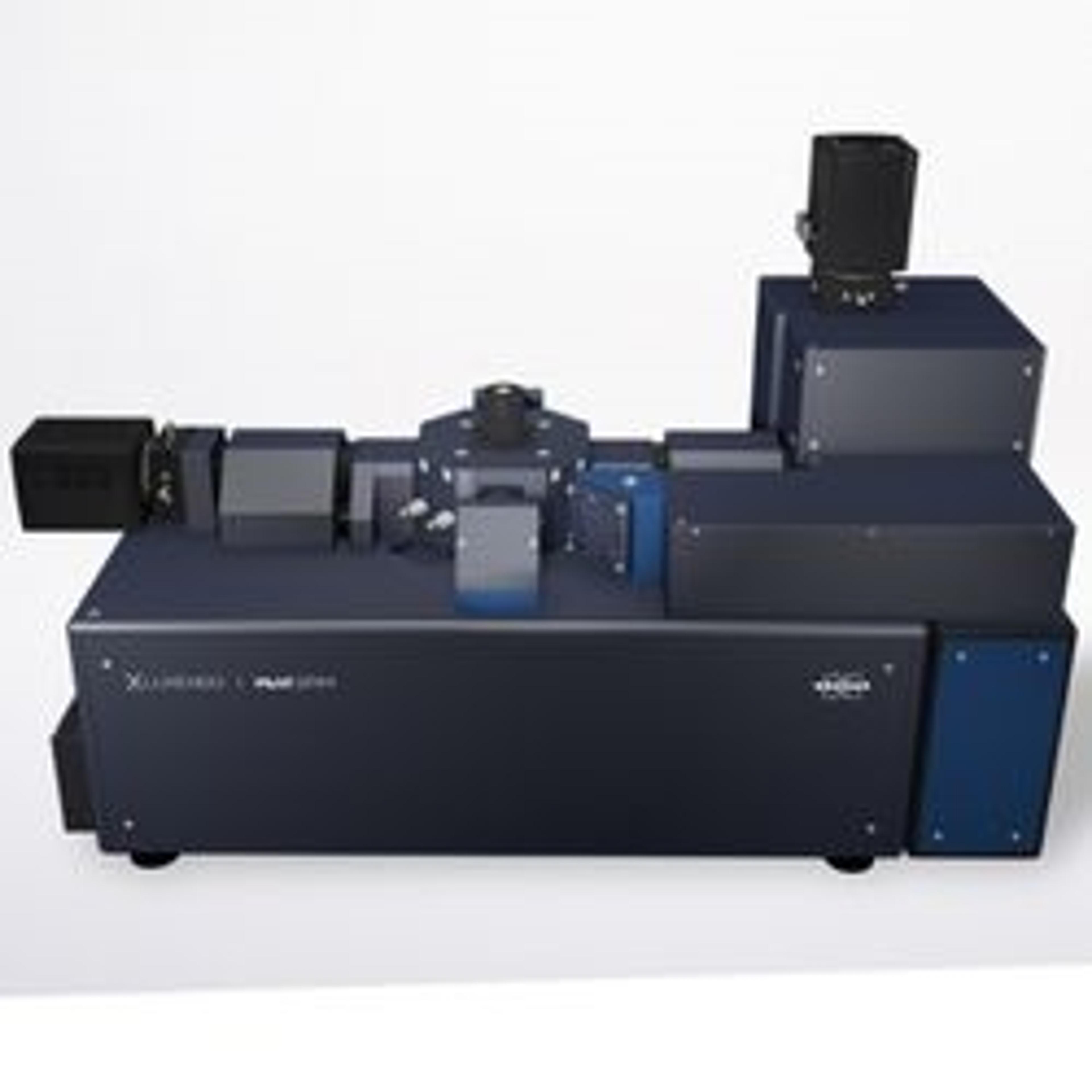
Aura® CL systems couples Fluorescent Membrane Microscopy and Backgrounded Membrane Imaging to help you understand what’s in your cell therapy. It detects, counts, sizes, and IDs cellular aggregates and subvisible particles and ID’s cellular from other particles without any machine learning so you can maximize the purity, safety, and efficacy of your therapeutic.
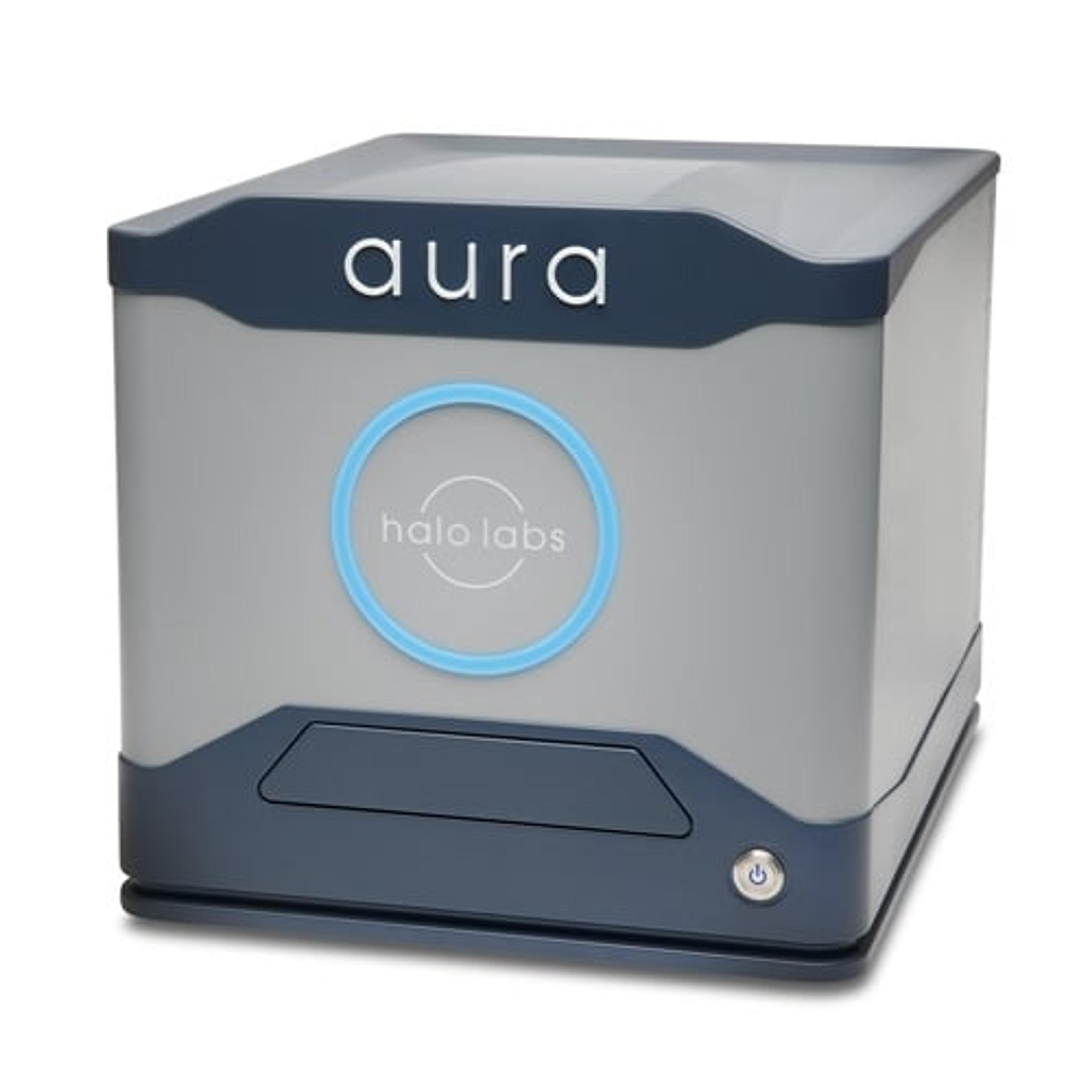
Aura® is the particle and aggregate detection system that combines Backgrounded Membrane Imaging which images 100% of your sample to give you count, size, and morphological information, with up to 2 channels of Fluorescence Membrane Microscopy. Identify if particles are protein or not with the first FMM channel and determine if your sample contains lipids, hydrophobic entities, or other aggregates of your choice with a second…
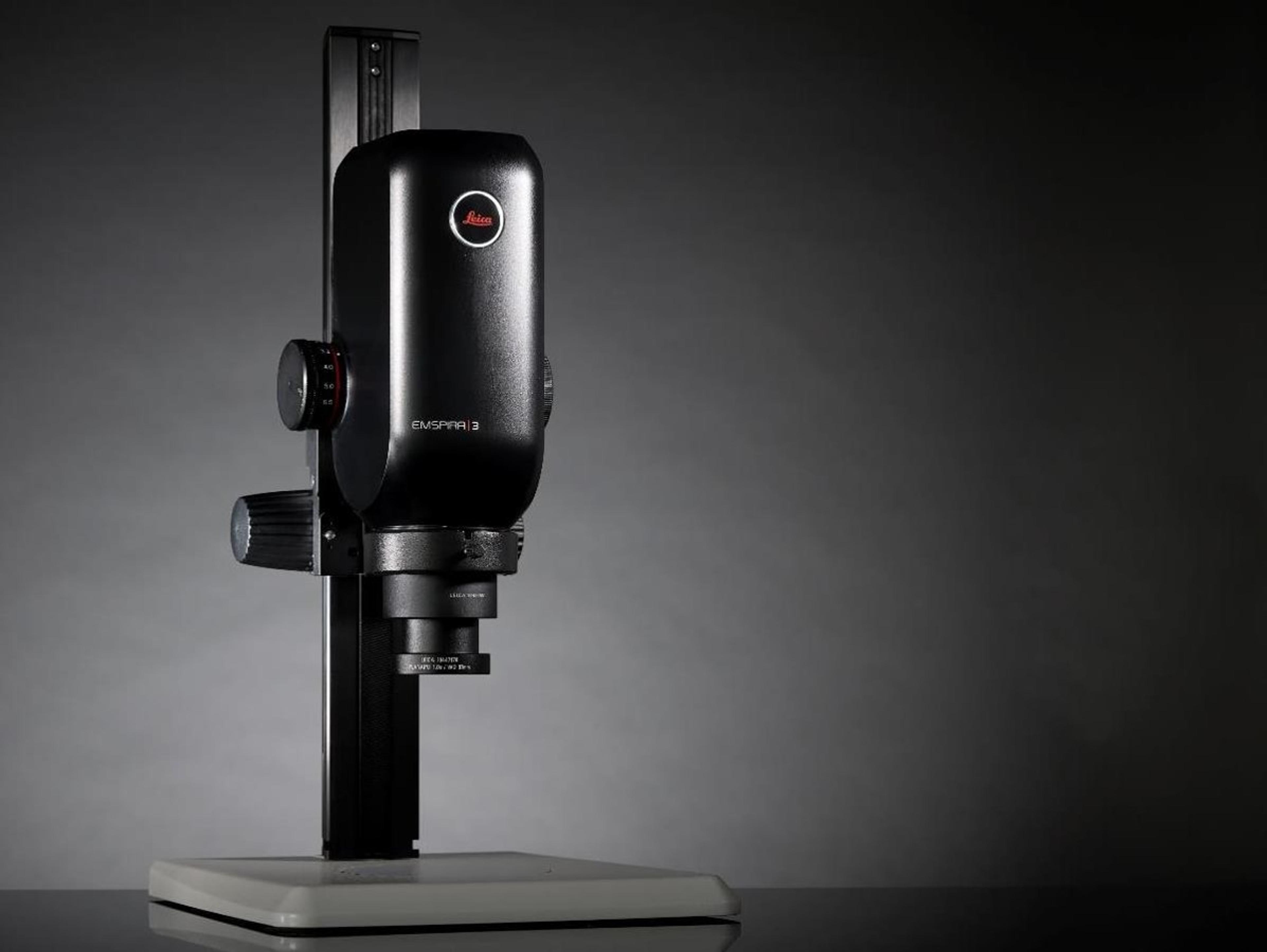
An all-in-one digital inspection solution from Leica Microsystems. The Emspira 3 digital microscope empowers users to streamline inspection processes, cover inspection needs flexibly, and work in a confident and reliable way, with a single system.
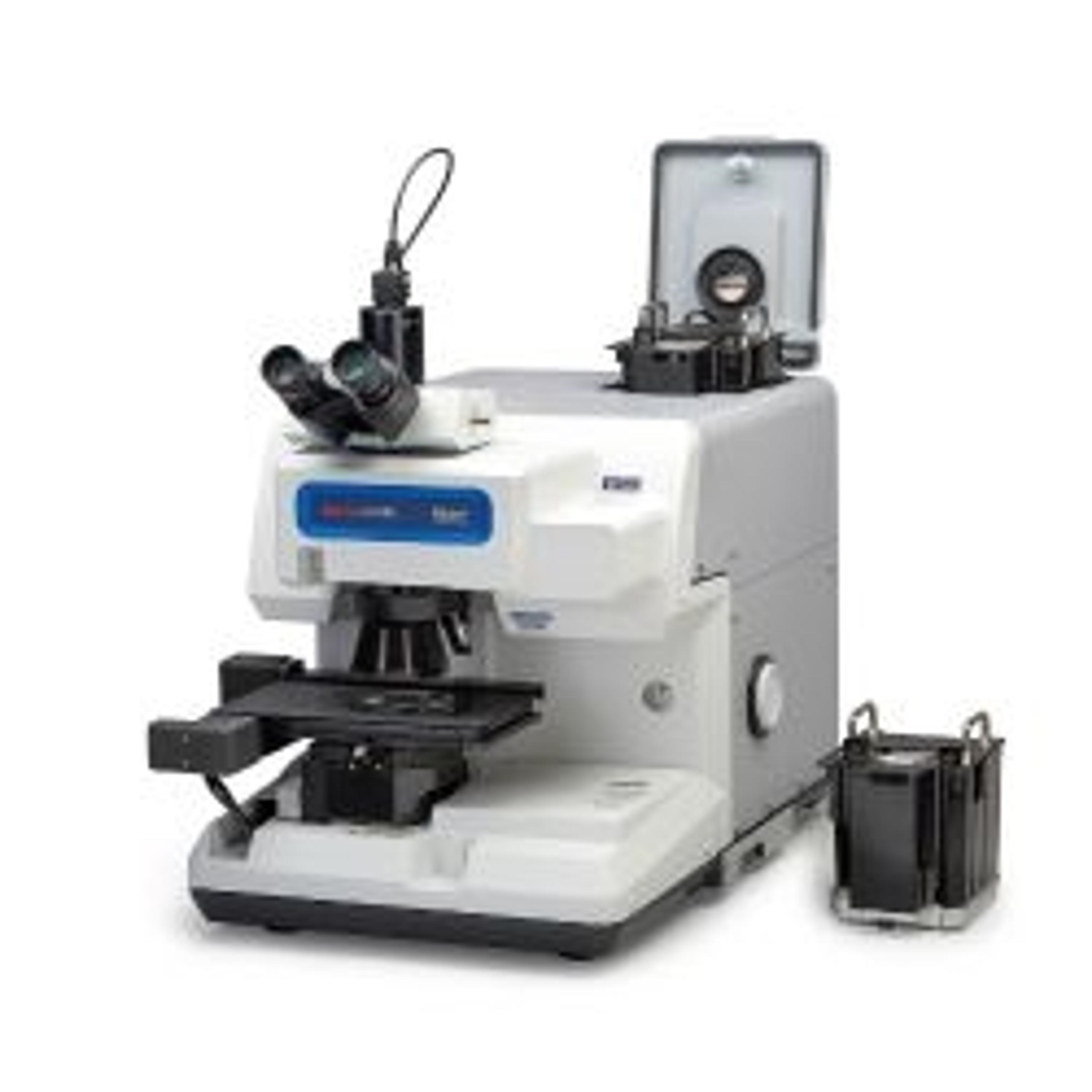
The newest generation InTouchScope™ SEM enhances productivity with Simple SEM automation. Automated image collection at multiple locations and conditions simplifies workflow for the most routine tasks. Embedded Signal Depth display enhances understanding of analytical spatial resolution. Seamless navigation from optical to SEM imaging, Live EDS (both spectrum and X-ray maps) and auto functions from alignment to focus produce…
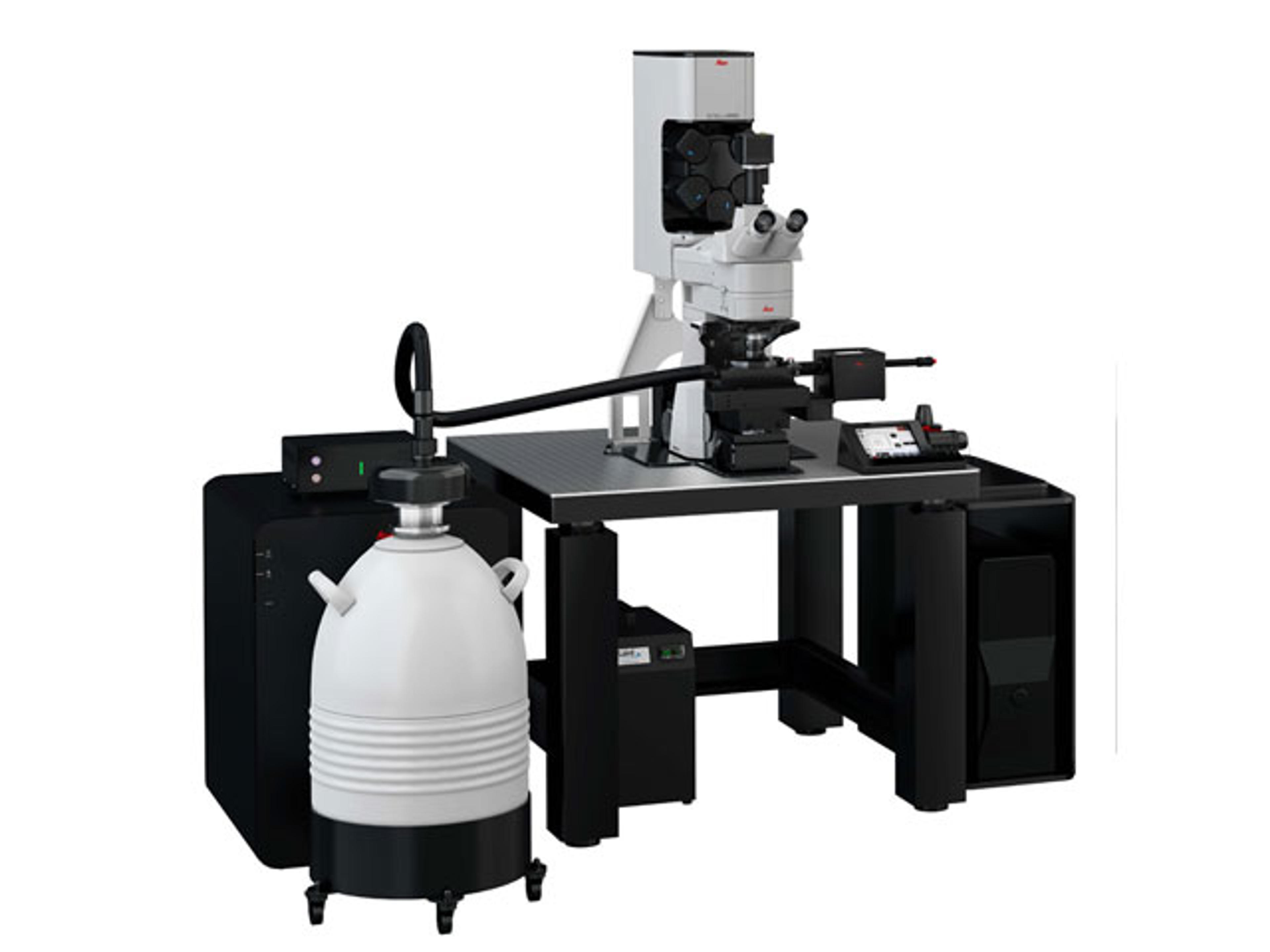
Mica is the world’s first imaging Microhub. More than a highly automated microscope, it unites widefield and confocal imaging in a sample protecting, incubating environment. With the simple push of a button, you have everything you need - all in one place - to supercharge fluorescence imaging workflows and get meaningful scientific results faster.
Leica's cryo-electron tomography workflow solution: boost your productivity with a seamless sample preparation workflow.
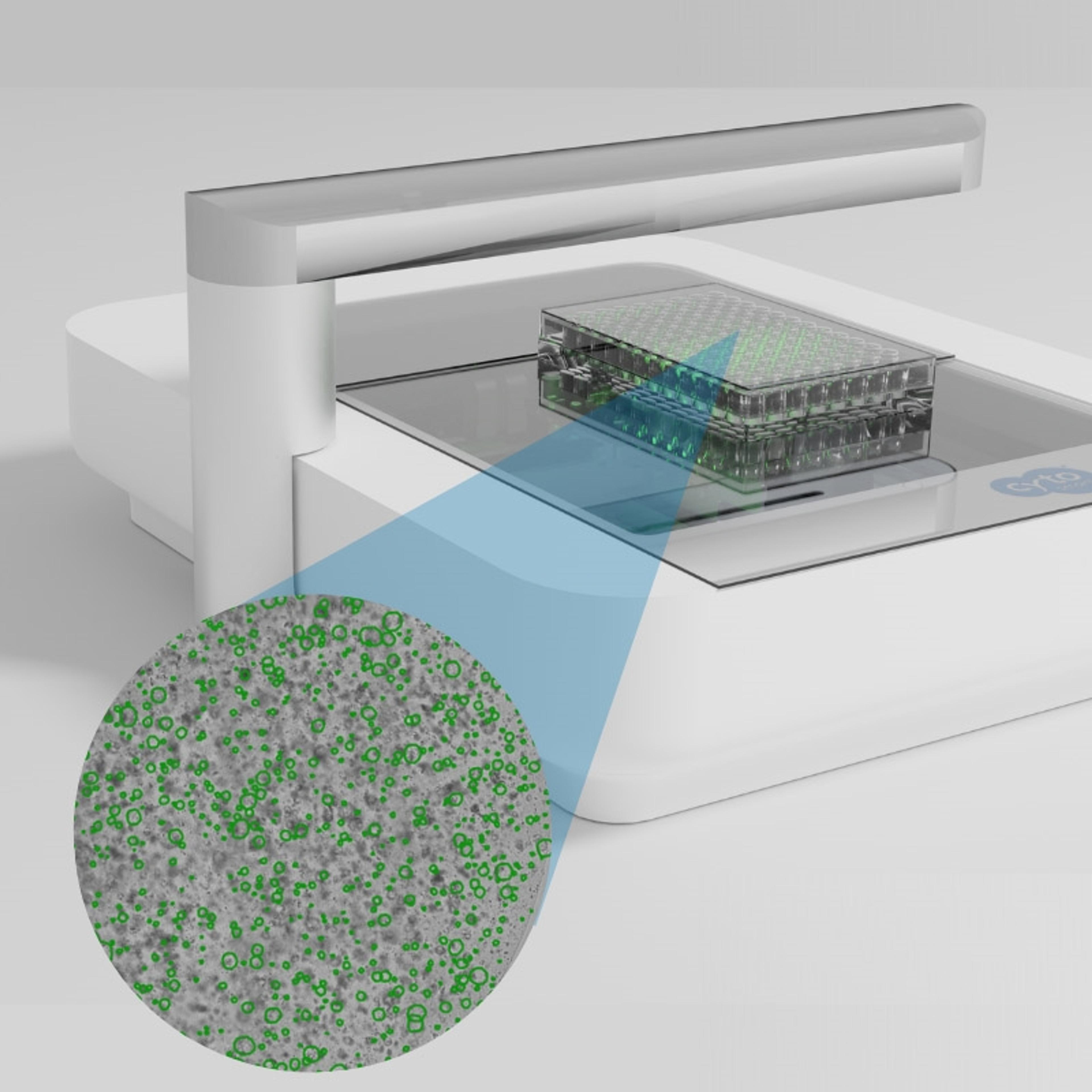
The Organoid Analysis Module for the CytoSMART Omni systems is an advanced machine-learning algorithm for robust organoid quantification, tracking, and analysis.
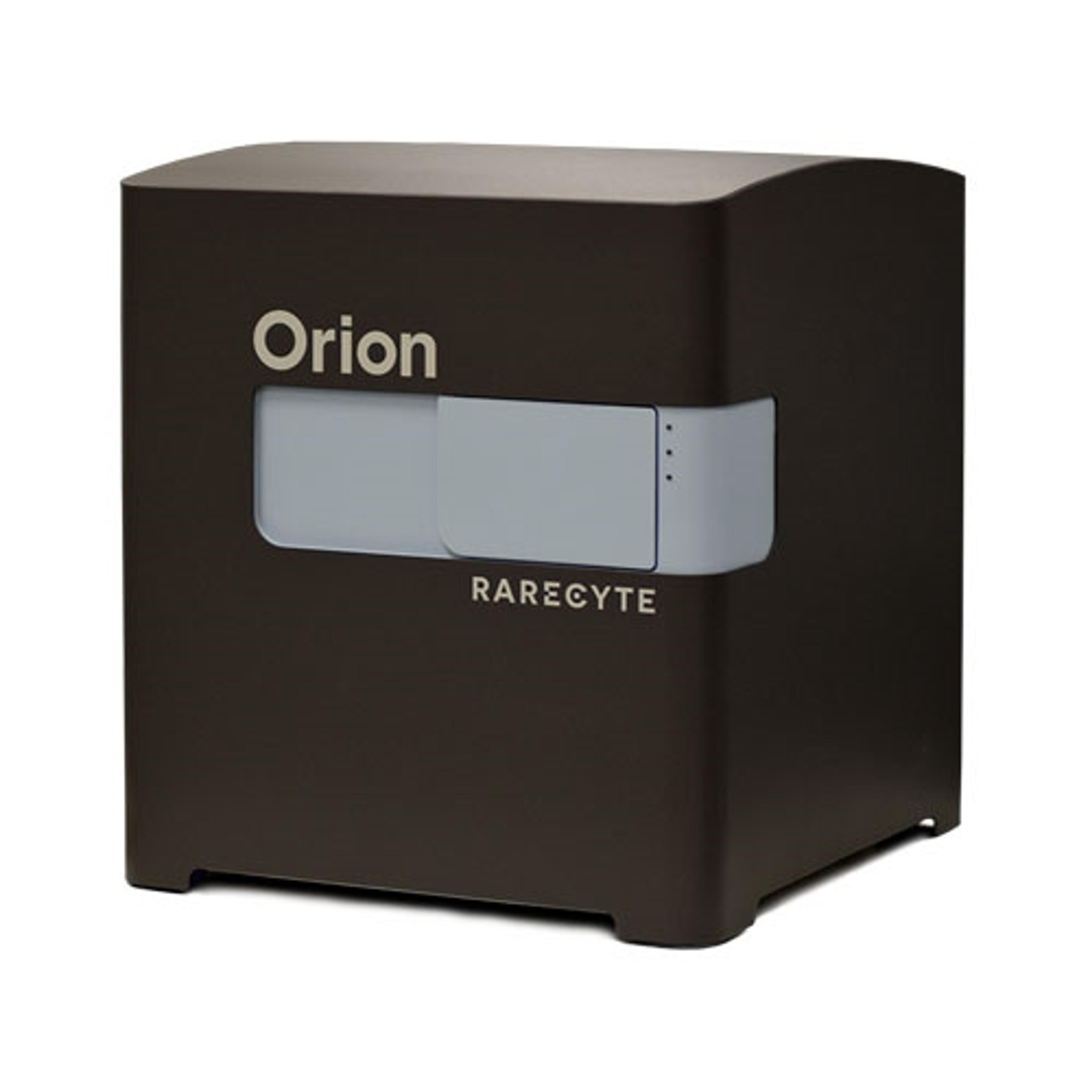
The Orion Platform delivers spatial biology and cellular phenotyping at a speed and resolution needed for translation from research to the clinic. It utilizes single round immunofluorescence staining and imaging of standard FFPE or fresh frozen tissue to deliver subcellular quantitation of biomarker targets across a whole slide in only a few hours. Combining speed and resolution with the additional benefits of industry-stan…
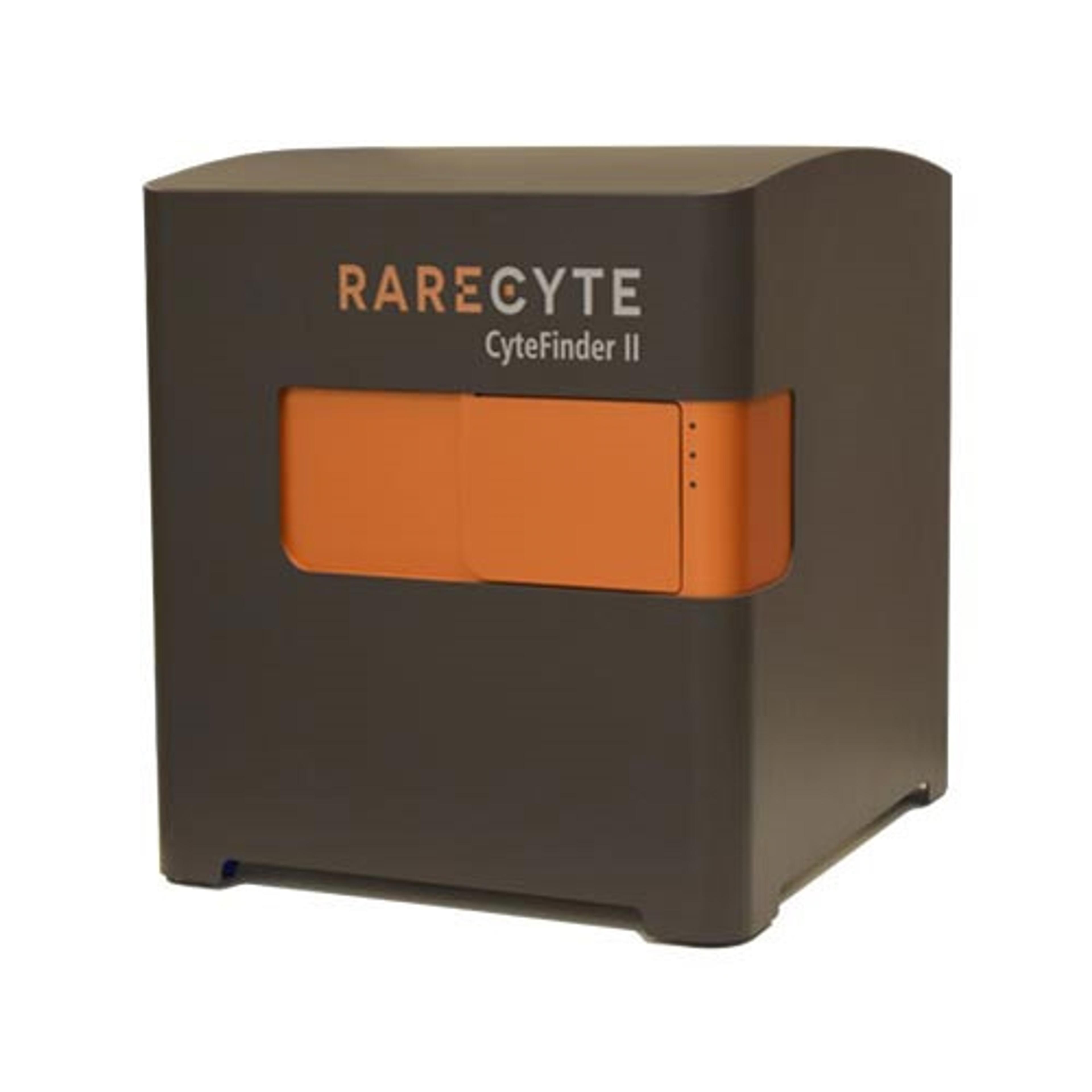
CyteFinder II Instruments are high speed, whole slide imaging systems with options for liquid biopsy analysis and multiplexed tissue imaging. Choose between the CyteFinder II with CytePicker® module for single cell or tissue micro-region retrieval, or the CyteFinder II HT for unattended scanning up to 80 slides at a time. High speed, multi-channel systems optimized for rare-cell detection and analysis Integrated m…
The Thermo Scientific™ Nicolet™ RaptIR+™ FTIR Microscope offers precision and agility to help streamline sample analysis by quickly generating actionable results. Homing in on the intricacies of a sample to find the answer you need is often a lengthy and difficult process. Any amount of time saved while searching for the solution makes a world of difference in delivering results.
Quickly identify unknown materials and contaminants that may affect product quality. The Thermo Scientific Nicolet iN5 FTIR Microscope provides rapid identification with point-and-shoot simplicity. The no-nonsense workflow requires only a few short steps. You can get answers in minutes—without the need for spectroscopy or microscopy expertise.
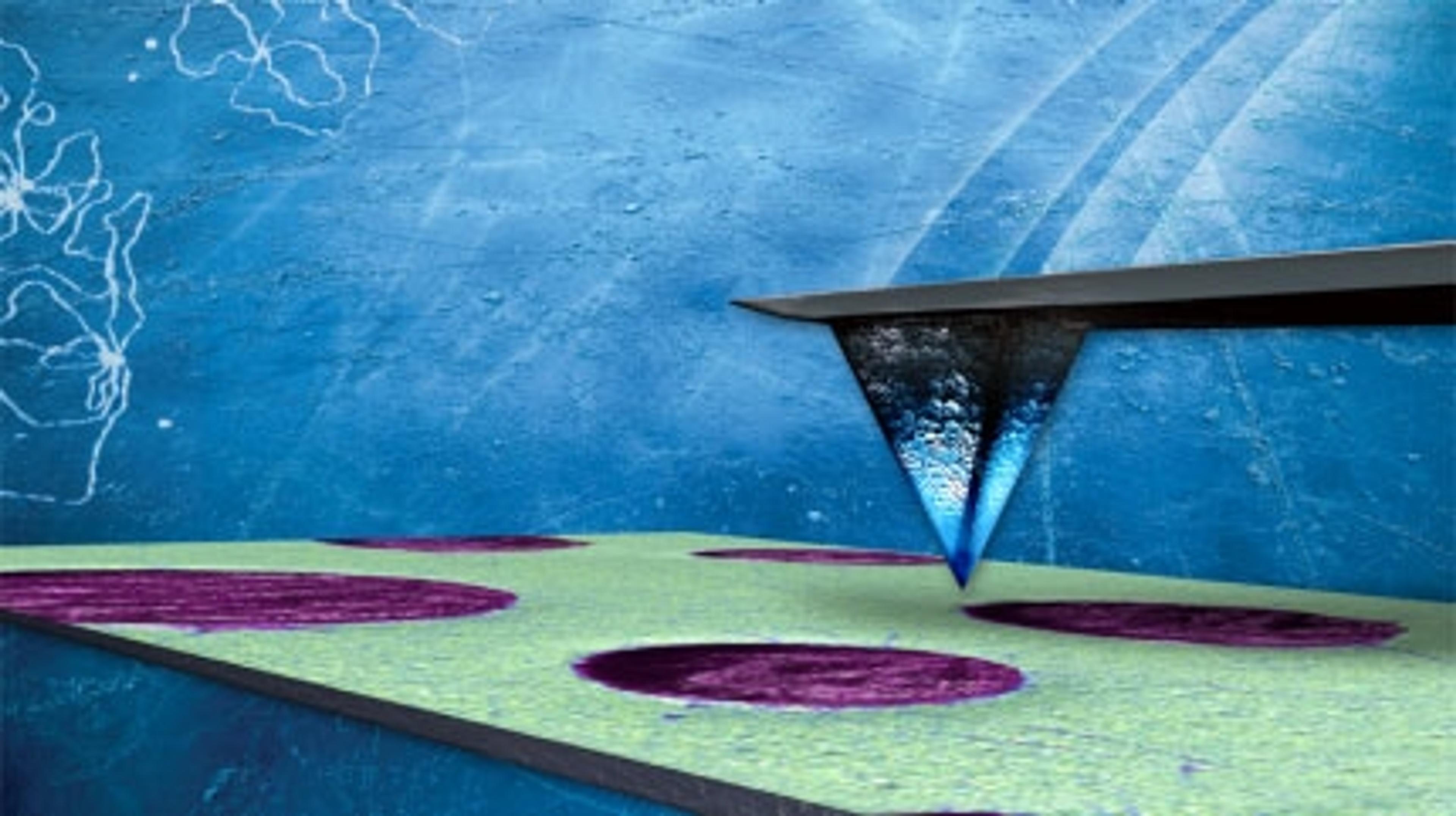
We offer a wide selection of AFM tips of various shapes, tip geometries, materials and coatings, spring constants, frequencies, etc. to fit your specific application. Whether we are manufacturing probes for the budget-conscious shopper or high-end performance applications, meeting a high standard of quality is priority to us. Our dedication to manufacturing probes, coupled with our expertise in AFM, ensures that we are unique…
The Thermo Scientific™ DXR3 Flex Raman Spectrometer is a research-grade Raman spectrometer specifically designed for integration with other analytical techniques. This compact spectrometer uses the same design and optics as advanced spectrometers in the Thermo Scientific DXR3 Raman Spectrometer research product family. Thus, it provides transportability and hyphenation without compromising data quality.
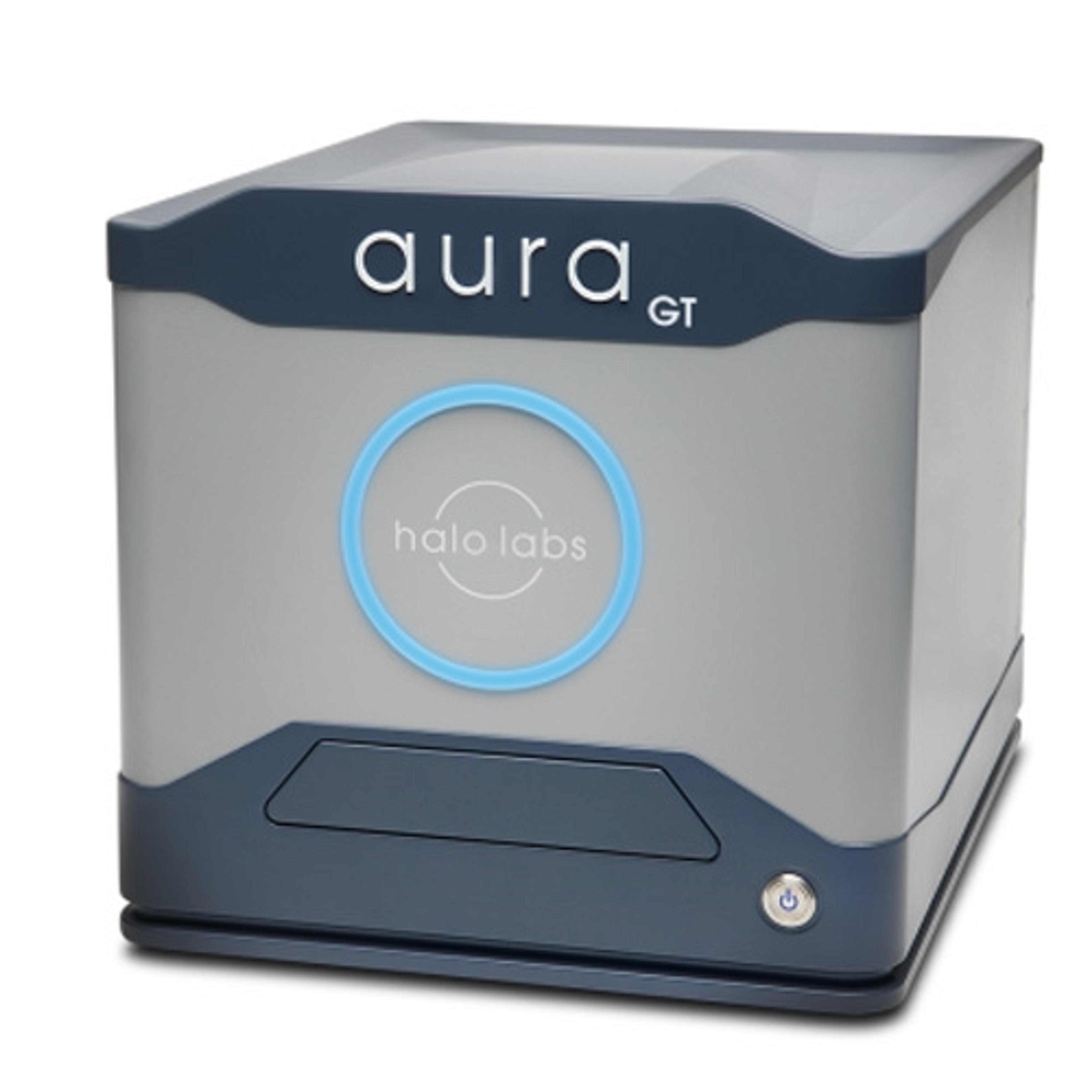
Aura® GT helps you realize the promise of gene therapies, without using a lot of your viral vector sample throughout the development process. Avoid conditions and formulations that facilitate viral vector aggregates, monitor DNA leakage, and ensure product quality with just 5 µL of sample per data point.
Aura® PTx gets you to your best formulation faster! Backgrounded Imaging (BMI) technology to give you accurate count, size, and morphology information about particles in your sample. Couple that with Fluorescence Membrane Microscopy (FMM), which gives you definitive ID of protein aggregates and an easy way to monitor polysorbate degradation.
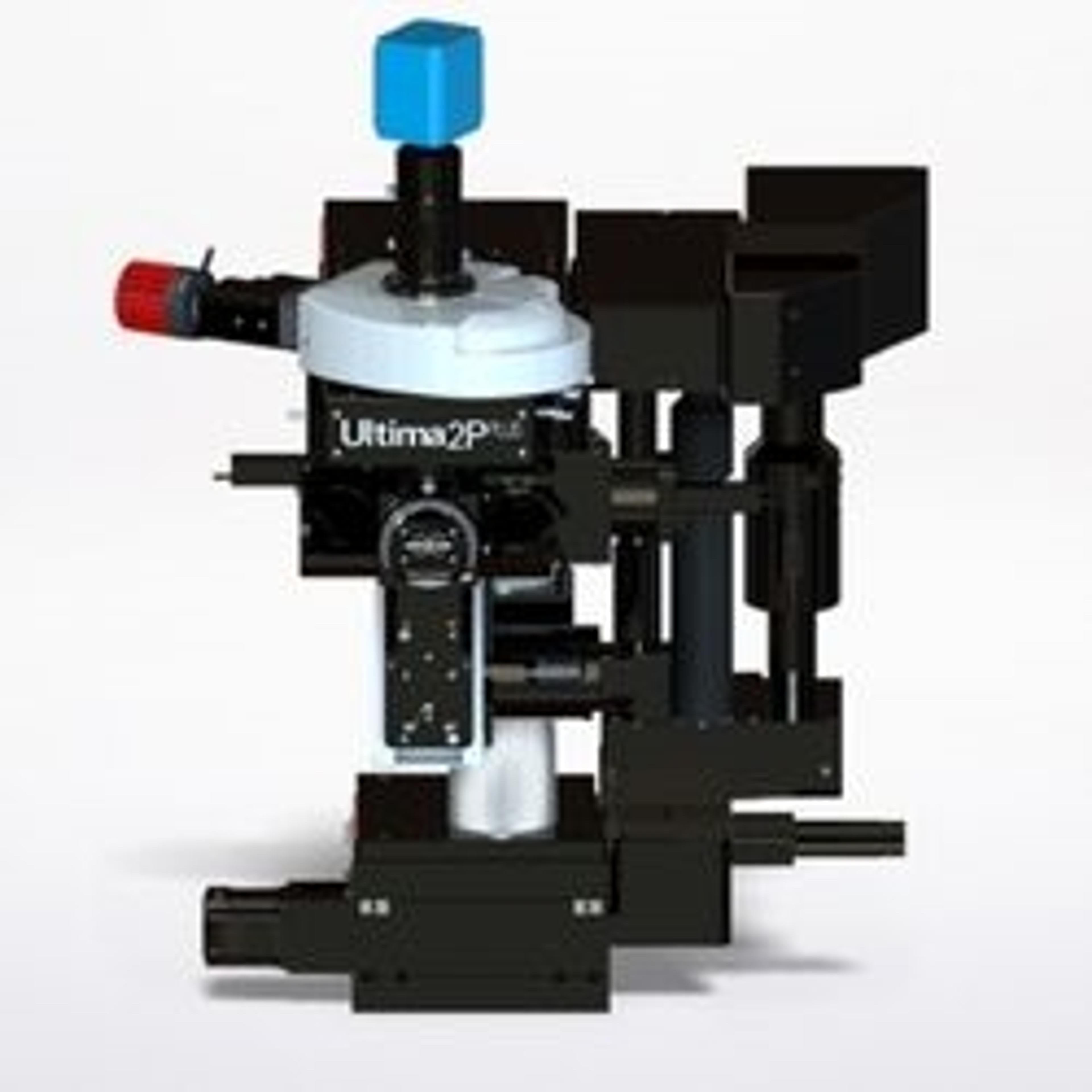
With new advances in field of view, sensitivity, wavelength, and sample accommodation, the Ultima 2Pplus delivers an ideal combination of flexibility, resolution, imaging depth and speed, allowing simultaneous imaging, stimulation and electrophysiology protocols with greater efficiency and effectivity. The system is designed specifically for intravital imaging, with fully motorized control of the objective X-Y-Z position, as…
The Vutara VXL comprehensive biological workstation for nanoscale biological imaging incorporates Bruker’s industry-leading single-molecule localization microscopy (SMLM) technology in a streamlined system with a compact footprint. The new system enables research on DNA, RNA, and proteins and supports advanced spatial biology research. When combined with Bruker’s unique microscope fluidics unit, Vutara VXL enables multiplexed…
Bruker microfluidics accessory units are designed to allow sequential labeling to be incorporated into localization microscopy protocols with the Vutara VXL single-molecule localization microscope. Bruker offers two units—PlexFlo and PlexFlo96. The units are controlled by Vutara's SRX software and allow flexible configuration of fluidics sequences that are run in conjunction with imaging cycles to allow users to label an unli…
Bruker's MuVi Selective-Plane Illumination Microscope (SPIM) is the fastest multi-angle view system on the market. MuVi incorporates years of Luxendo light-sheet technology innovations to provide high-speed volumetric acquisition for analyzing dynamic processes in delicate live specimens, as well as offering compatibility with any clearing method. A variety of other advanced hardware and software integrations can be easily co…
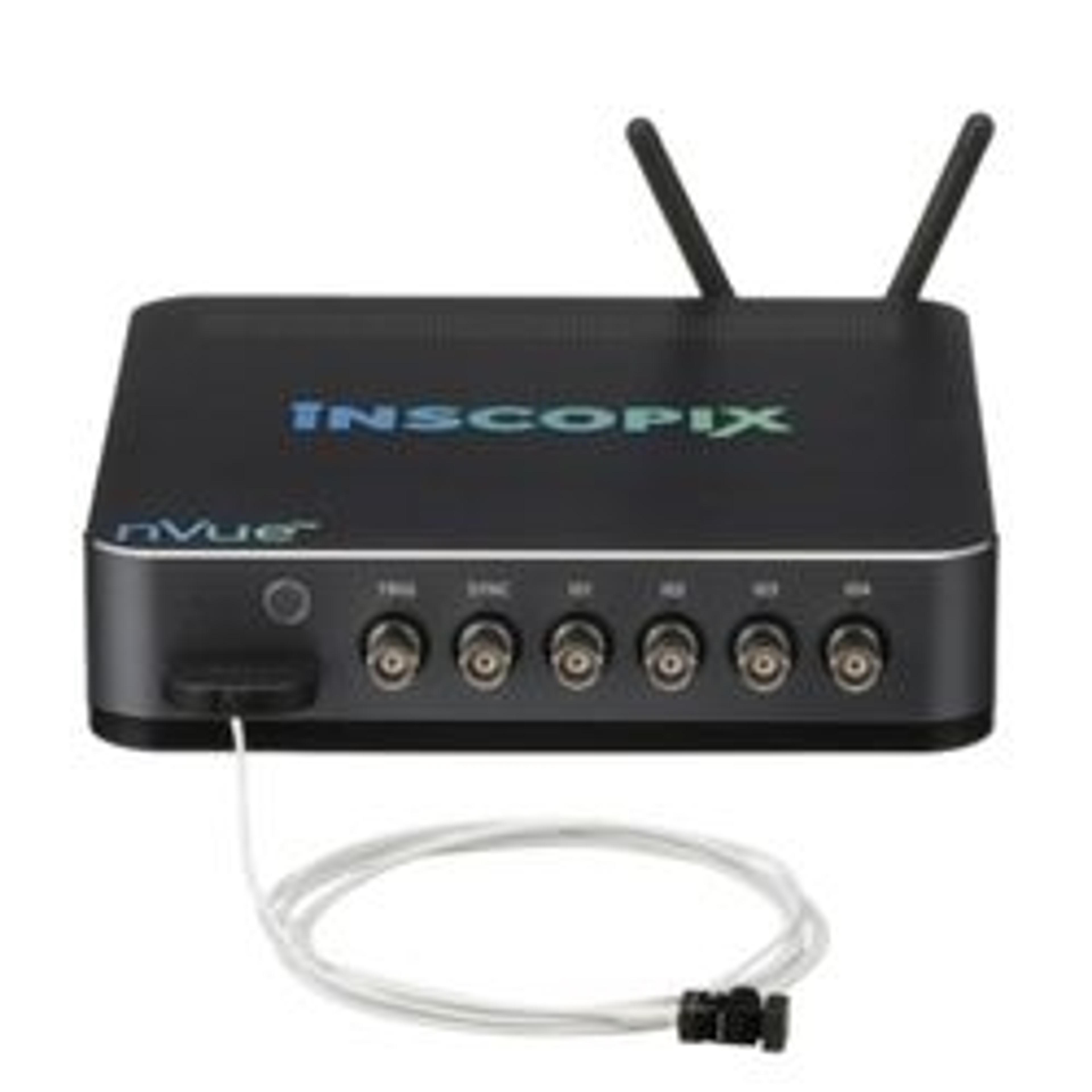
Inscopix’s nVue system is a complete miniscope platform able to register dual-color calcium signals from two cellular populations simultaneously with single-cell resolution in freely behaving animals. A streamlined workflow supports static and dynamic cell imaging, blood flow imaging, or genetically encoded optical neurotransmitter sensors, all within the same field of view. Furthermore, this technology is supported by multim…
The Chromatone system offers an integrated, multi-dimensional approach for simultaneous calcium imaging and optogenetic stimulation in freely behaving animals. It is a small and ultralight fiber-bundle technology that delivers simultaneous multi-region neural imaging and optogenetic manipulation in up to four regions for flexible investigation of the central nervous system or spinal cord with single-neuron resolution — enabli…