Light Microscopy Products & Reviews
25
Selected Filters:

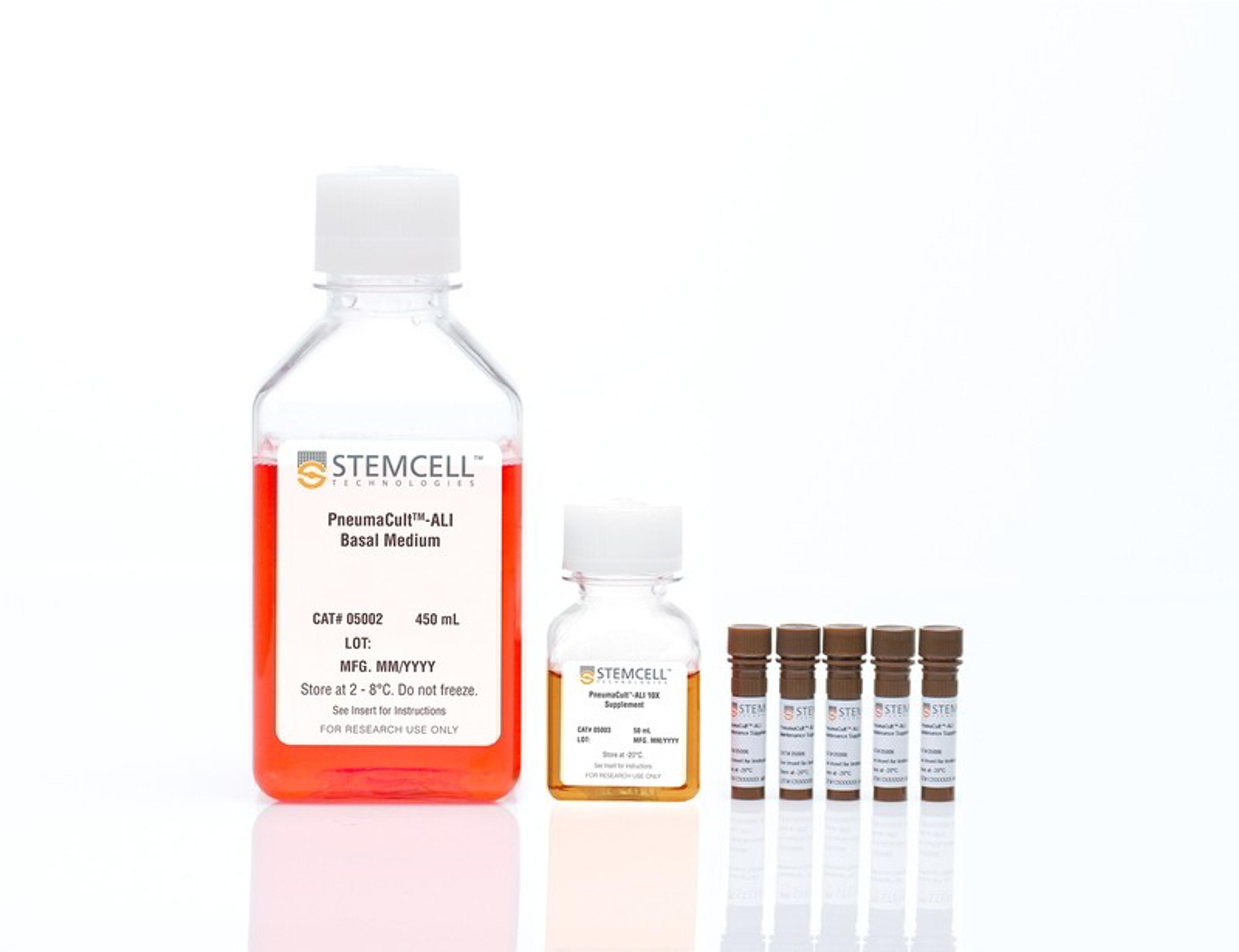
PneumaCult™-ALI Medium
STEMCELL Technologies Inc.Serum- and BPE-free medium for human airway epithelial cells cultured at the air-liquid interface or as airway organoids
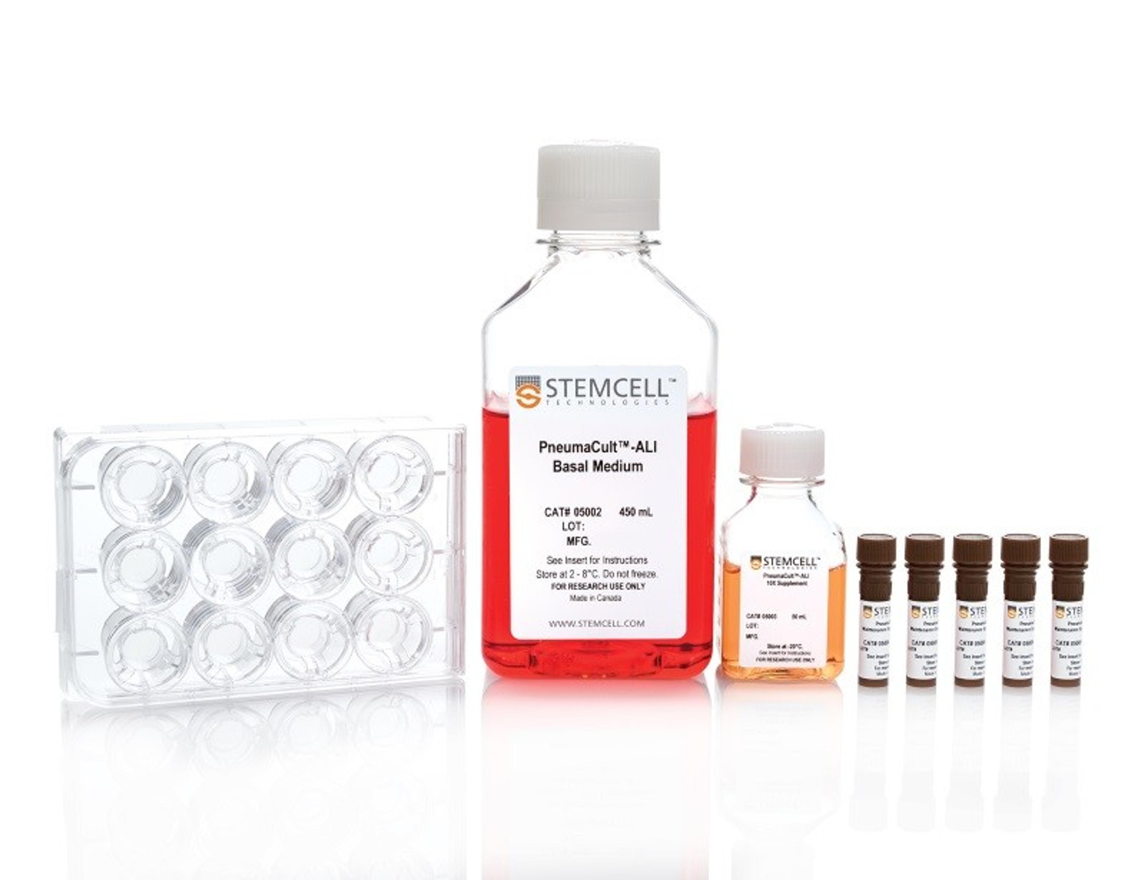
PneumaCult™-ALI Medium with 12 mm Transwell® Inserts
STEMCELL Technologies Inc.Serum- and BPE-free medium for human airway epithelial cells cultured at the air-liquid interface or as airway organoids

PneumaCult™-ALI Medium with 6.5 mm Transwell® Inserts
STEMCELL Technologies Inc.Serum- and BPE-free medium for human airway epithelial cells cultured at the air-liquid interface or as airway organoids

PneumaCult™-Ex Plus Medium
STEMCELL Technologies Inc.Serum- and BPE-free medium for expansion of primary human airway epithelial cells

SensoScope® Brightfield
Miltenyi Imaging GmbHPremium digital microscope for live imaging, scanning and real-time diagnostics of multiple slides

Thermo Scientific™ HCS Studio Cell Analysis Software
Thermo Fisher ScientificIntuitive interfaces, intelligent design and powerful image analysis tools leading to meaningful dynamic measurements at the cell-, well- and field-level.
Invitrogen™ Celleste Image Analysis Software
Thermo Fisher ScientificImage processing, manual and automatic measurements over multiple channels, to segmentation and classification tools that help you transform images into quantitative data

FlowCam LO
Yokogawa Fluid Imaging Technologies, IncInnovative particle characterization with FlowCam® LO combines flow imaging microscopy (FIM) and light obscuration (LO) into a single analytical solution. Beyond the compendial light obscuration method to fulfill USP <787> and <788> requirements, flow imaging microscopy provides an orthogonal method for quality control of subvisible particulate matter.

Provi™ CM20 Incubation Monitoring System
EVIDENTThe CM20 system provides quantitative data remotely - place the head and your cell cultures in the incubator, and the system will periodically scan it, count the number of cells, and determine confluency. The data are wirelessly communicated to a PC or a tablet through an optional router, so you can monitor your cultures' progress without entering a clean room.

ibidi Stage Top Incubation System, with CO2 and O2 Control, for Live Cell Imaging
ibidi GmbHEasy setup directly on your microscope: perform live cell imaging in dishes, slides, or multiwell plates. The ibidi Stage Top Incubation Systems fit every standard inverted microscope and include CO 2 and O 2 control as well as actively controlled humidity. They are ideal for all live cell imaging applications and available for single slides and dishes as well as for multiwell plates.

µ-Dish 35 mm, high
ibidi GmbHUse this versatile 35 mm imaging Dish for high-end microscopy through the #1.5 coverslip bottom.
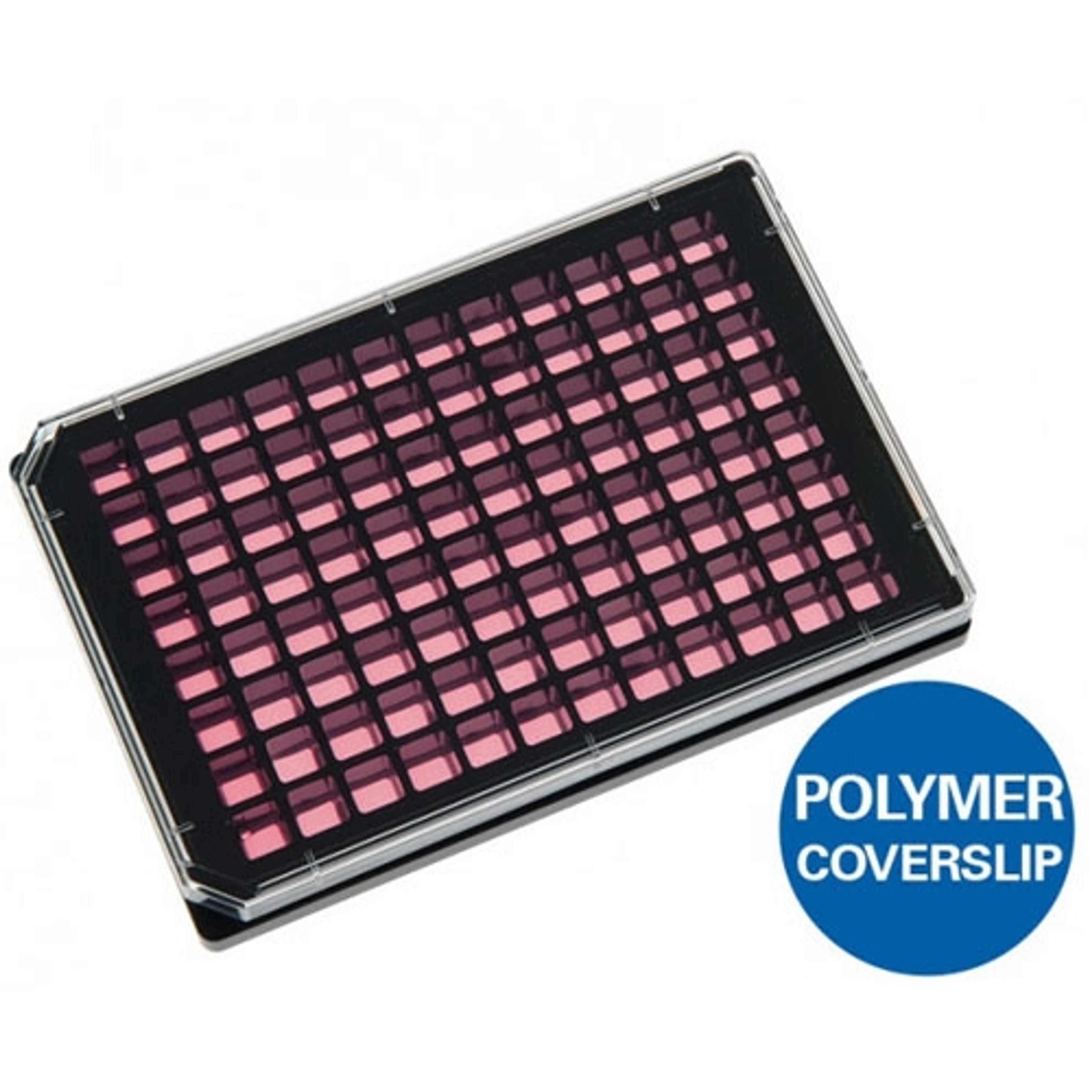
µ-Plate 96 Well Black
ibidi GmbHUse this Multiwell plate with black walls, square wells, and a flat and clear coverslip bottom (#1.5 ibidi Polymer Coverslip) for high-throughput applications in cell-based assays. Also available with 24 wells.
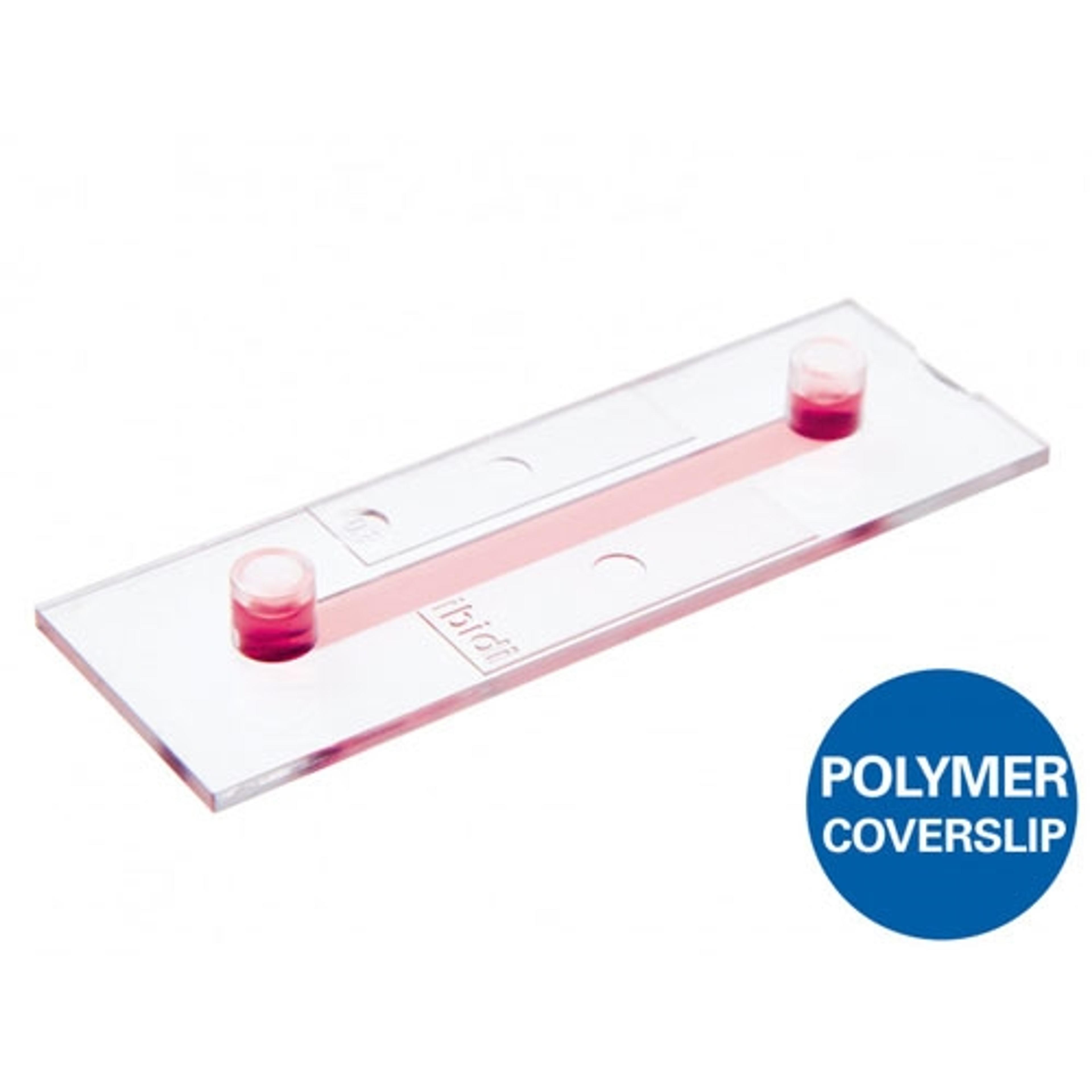
µ-Slide I Luer
ibidi GmbHUse this channel microscopy slide with a coverslip bottom (#1.5 ibidi Polymer or #1.5H glass) for immunofluorescence, cell culture under flow, live cell imaging, and high resolution microscopy on inverted microscopes. Available in various channel heights.
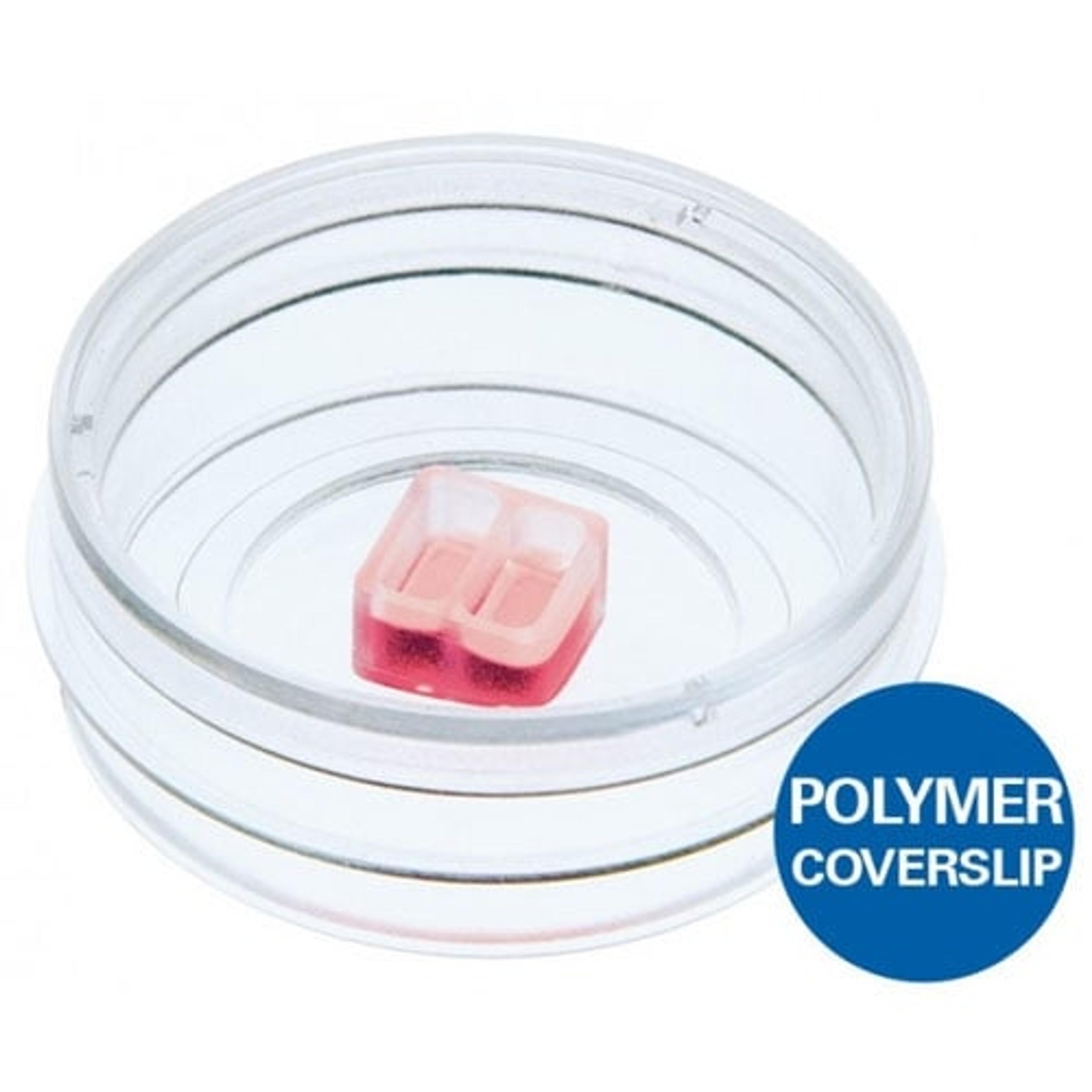
Culture-Insert 2 Well | 3 Well | 4 Well
ibidi GmbHUse these silicone inserts with a defined cell-free gap for wound healing assays, migration assays, 2D invasion assays, and co-cultivation of cells. Available with 2, 3, or 4 wells.

µ-Slide Angiogenesis
ibidi GmbHUse this slide to investigate angiogenesis in tube formation assays. Also ideal for 3D cell culture and immunofluorescence staining. Also available in a 96 well format.

µ-Slide Chemotaxis
ibidi GmbHInvestigate chemotaxis of fast or slow migrating adherent cells and non-adherent cells in 2D or 3D gel matrices.

Aivia AI Microscopy Image Analysis Software
Leica Microsystems EuropeAccess the future of AI microscopy. Using state-of-the-art, AI-first software architecture, Aivia is a uniquely innovative and complete 2-to-5D image visualization, analysis and interpretation platform designed for the reliable processing and reconstruction of highly complex images in just minutes. Aivia provides powerful 2D & 3D spatial relational analysis for objects across diverse types, morphological complexities, and hie…

Cell DIVE Multiplexed Imaging Solution
Leica Microsystems EuropeCell DIVE multiplexed imaging is an antibody-based hyperplexed technique addressing spatial cell biology and function within the tumor microenvironment.

STELLARIS FALCON FLIM Microscope
Leica Microsystems EuropeSTELLARIS 8 FALCON (FAst Lifetime CONtrast) is the future of functional imaging. Harness the power of fluorescence lifetime to investigate cellular physiology and explore dynamics in living cells.

STELLARIS DIVE Multiphoton Microscope
Leica Microsystems EuropeDIVE and STELLARIS have now merged to provide you with the power of flexible multicolor multiphoton imaging.

STELLARIS DLS Digital Light Sheet Microscope
Leica Microsystems EuropeSTELLARIS LightSheet (DLS) unites in one place a confocal system and a light sheet microscope – a unique combination aimed to make your research more versatile. DLS and STELLARIS enable even gentler imaging giving you the ability to perform fast and gentle volumetric light sheet imaging and to improve your live cell imaging applications.
STELLARIS Coherent Raman Scattering (CRS) Microscope
Leica Microsystems EuropeWhen you need to study structures that cannot be visualized with traditional fluorescent microscopy methods, the STELLARIS 8 Coherent Raman Scattering (CRS) microscope enables you to implement label-free chemical imaging into your workflow to answer those challenging research questions.
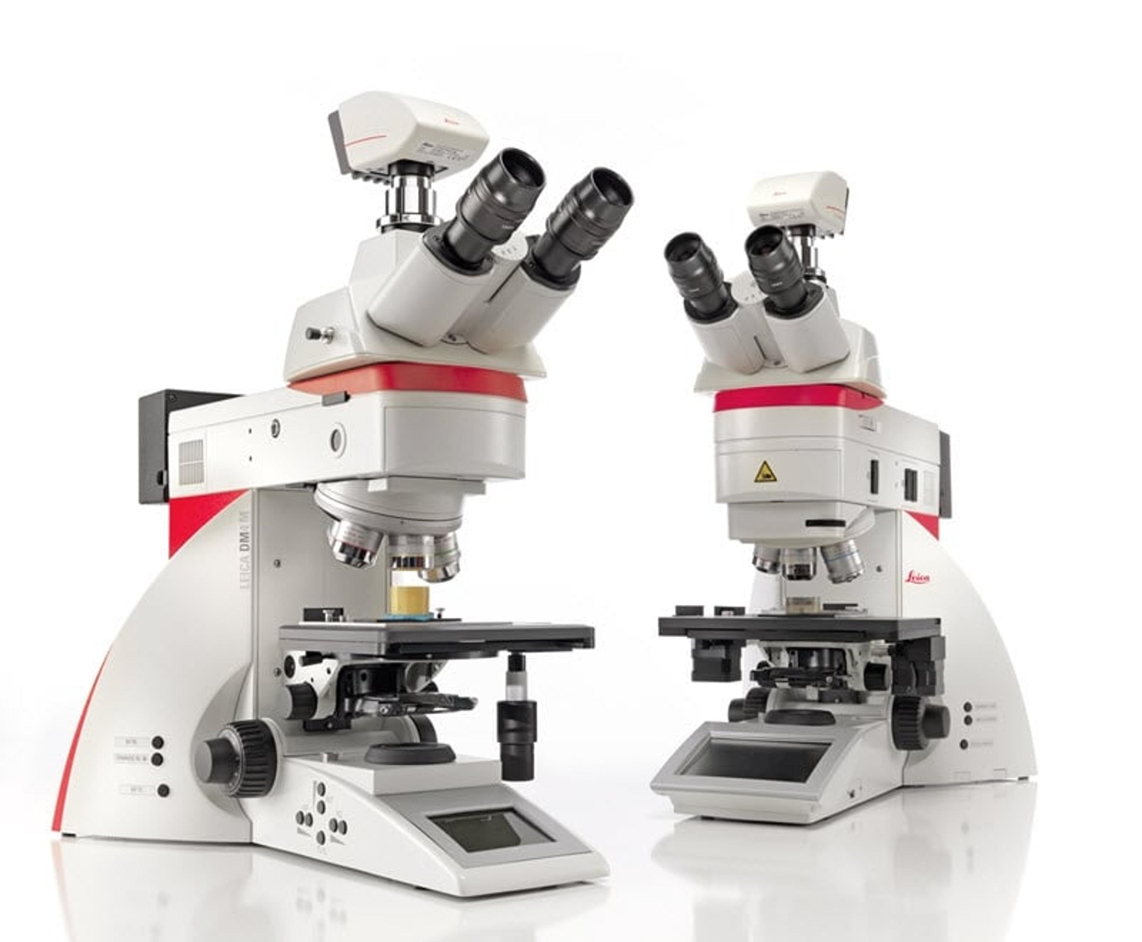
DM4 M & DM6 M Upright Materials Microscopes
Leica Microsystems EuropeDo you need to image, measure, and analyze similar features across many samples and materials science and analysis? The Leica DM4 M and Leica DM6 M are the Industrial Microscopes for you, whether microscopy novice or professional.