Reformatting Services
ASDI BioSciencesASDI will reformat your library for automation compliance, transferring compounds from an old format to a new standard format capatable with Automated Sample Retrieval Systems (ASRS).
Lab informatics harnesses cutting-edge technologies to manage, analyze, and interpret the vast amounts of data generated in lab settings. It encompasses the use of advanced software systems, including laboratory information management systems (LIMS), electronic laboratory notebooks (ELN), and scientific data management systems (SDMS), to streamline workflows and facilitate collaboration. Increasingly, lab informatics integrates machine learning (ML) and artificial intelligence (AI) algorithms to automate data analysis, predict trends, and optimize experimental processes. Explore how lab informatics empowers researchers and scientists to derive actionable insights, accelerate discoveries, and drive advancements across diverse scientific fields.
ASDI will reformat your library for automation compliance, transferring compounds from an old format to a new standard format capatable with Automated Sample Retrieval Systems (ASRS).
ASDI provides weight capture of empty vials which can be bar-coded prior to taring. ASDI can supply these bar-codes or use a bar-code supplied by a client.
ASDI will identify decomposed samples in your library, confirm chemical synthesis, or validate the quality of screening samples purchased from 3rd party vendors. NMR LC-MS GC HPLC
ASDI maintains a state of the art liquid handling suite that manipulates 10's of thousands of compounds per week, formatting stock libraries or your newly purchased samples into industry standard plates. ASDI may also condense existing plate libraries into 384-well plates or another compression standard. We follow rigously validated SOP's, yet remain flexible to meet your custom requirements. Sample Solvating Condensing…
Revolutionary 2DGE Software Analysis of single 2D gels - Dymension 1 is ideal for users looking for software to analyse spots between samples containing one 2D gel only. It offers a range of automatic image correction features and methods for resolving and analysing spots on single gels. The software can be easily upgraded to Dymension 2 or 3 if the proteomics needs of the laboratory change.
Cellomics Store offers an open, enterprise-ready, relational database solution, for the management of data generated by today’s high content screening (HCS) instrumentation and image analysis tools. HCS technologies rapidly generate large volumes of data and images which are automatically organized by Cellomics Store for easy user access.Store is field proven and its ability to automatically link images, derived data (e.g. res…
CellSpace® Knowledge Miner is a revolutionary web based tool for searching the scientific literature. Rapidly, visually, discover ranked relationships from the literature to scientifically qualify targets, genes, proteins and screening results. CellSpace continuously monitors relationships from 650+ journals in MedLine and will email you if relationships of interest change. Academic, concurrent and volume licensing is ava…
vHCS™ Discovery ToolBox software, provides a complete set of powerful tools to relate, manage, analyze, visualize and communicate High Content Screening (HCS) and High Content Analysis (HCA) images, data, information and results. Use our powerful tools to drive your HCS and HCA discovery efforts, through improved productivity and rapid decision making. Use the ToolBox (with Cellomics® Store) to globally share HCS data as well…
vHCS:View Module Desktop PC software is avalable separately from the full vHCS™ Discovery ToolBox. vHCS:View provides, Rich visualization of HCS data, allowing users to see spatial, temporal, morphological and phenotypic changes at the cell levelInteractive visual analysis of multi-feature, cell-based data to the image levelLink to Spotfire® DecisionSite™ for deeper visualization and data miningMulti-plate, multi-well, multi-…
Cellomics® Content Addressed Storage (CAS) Module for EMC Centera™ is an HCS Applications Server plug-in that provides an interface to the EMC Centera content addressed storage system. Centera is EMC’s newest storage platform specifically designed to work with large volumes of fixed content information, such as the many terabytes of images generated by HCS instrumentation. The CAS Module allows HCS image data to be seamlessly…
Cellomics HCS Applications Server (HCS-AS) is the middleware framework in Cellomics' High Content Informatics (HCiTM ) software platform for High Content Screening ,High Content Analysis and cellular discovery.The HCS-AS performs a critical role in the overall HCS process, providing the business level logic for HCS and high content analysis as well as the data access from instruments and vHCS Discovery ToolBox clients to Cello…
Cellomics® High Content Informatics (HCi™) is the software platform of choice for High Content Screening(HCS) and High Content Analysis (HCA). The simple goal of HCi is to provide life scientists and life science IT professionals with an enterprise class, highly robust, open, industry standard software infrastructure to maximize the value of HCS. HCi software transforms images into information and then into knowledge, in orde…
The Micronucleus V2 BioApplication is a powerful image analysis algorithm for identifying and quantitating the rare event of micronucleus formation as a measure of genotoxicity. Because it can screen a 96-well plate in under 8 hours, it enables screening for genotoxic compounds in an automated, objective manner. This application has been biologically validated, enhancing confidence in your image-based data. Available for th…
Molecular Translocation V2 is an advanced image analysis application for quantitating simultaneous cytoplasm-to-nucleus translocation of multiple targets. It features subpopulation selection and cross-correlated responder analysis of up to 5 distinct targets, allowing study of signaling pathways. This application has been biologically validated, enhancing confidence in your image-based data. Available for the Cellomics Ar…
Morphology Explorer V2 is a powerful general-purpose image analysis application for measuring size, shape, and distribution of cells, colonies, and subcellular components. It performs sophisticated calculations in a turn-key, automated manner allowing higher throughput of complex cell-based assays. This application has been biologically validated, enhancing confidence in your image-based data. Available for the Cellomics…
Cell Health Profiling V2 is a flexible image analysis application employing advanced cell-level logic to detect and classify cell health-related events. It analyzes patterns of cellular fluorescent labeling that can be used to classify cells as undergoing apoptotic, necrotic, or other cell death mechanisms. This application has been biologically validated, enhancing confidence in your image-based data. Available for the Cel…
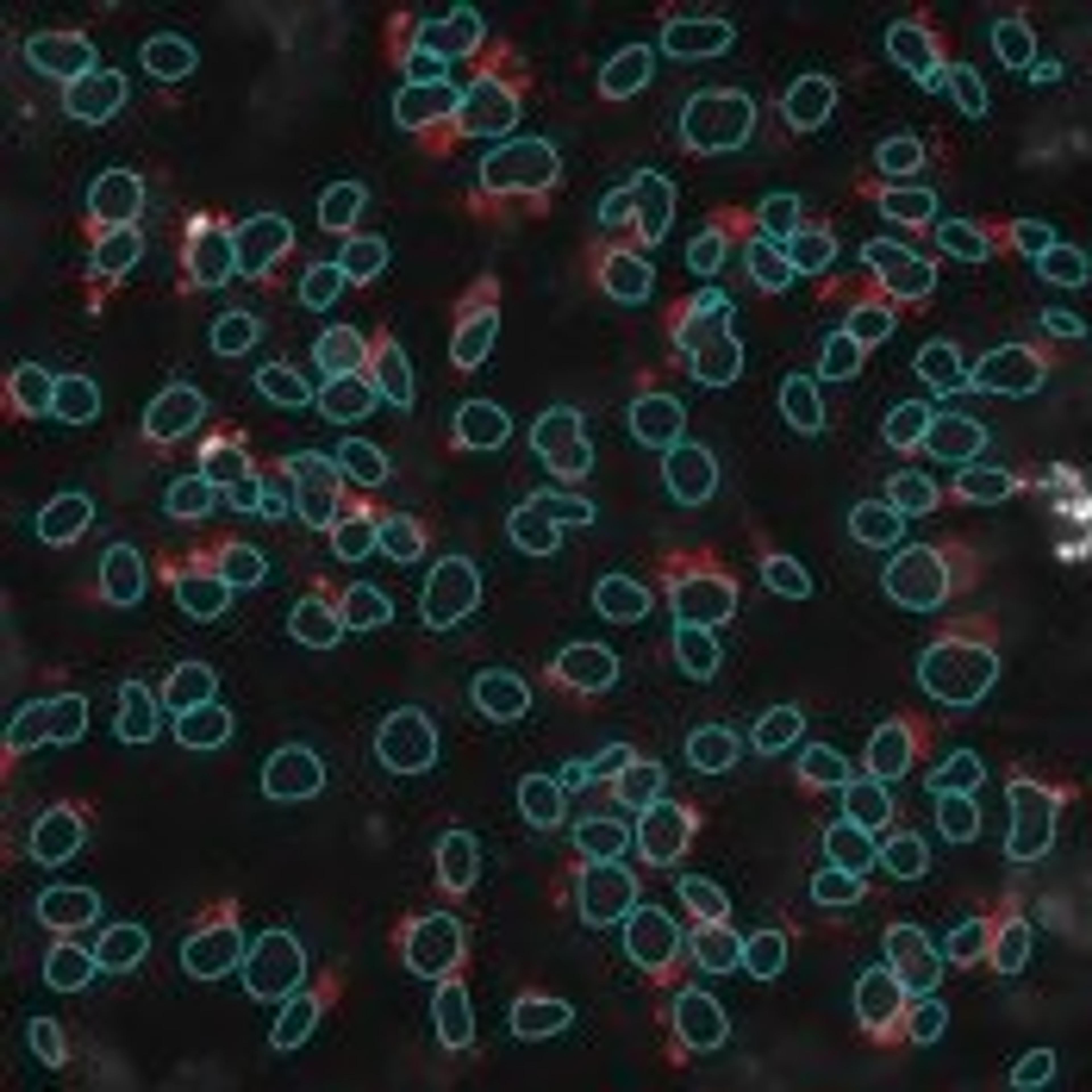
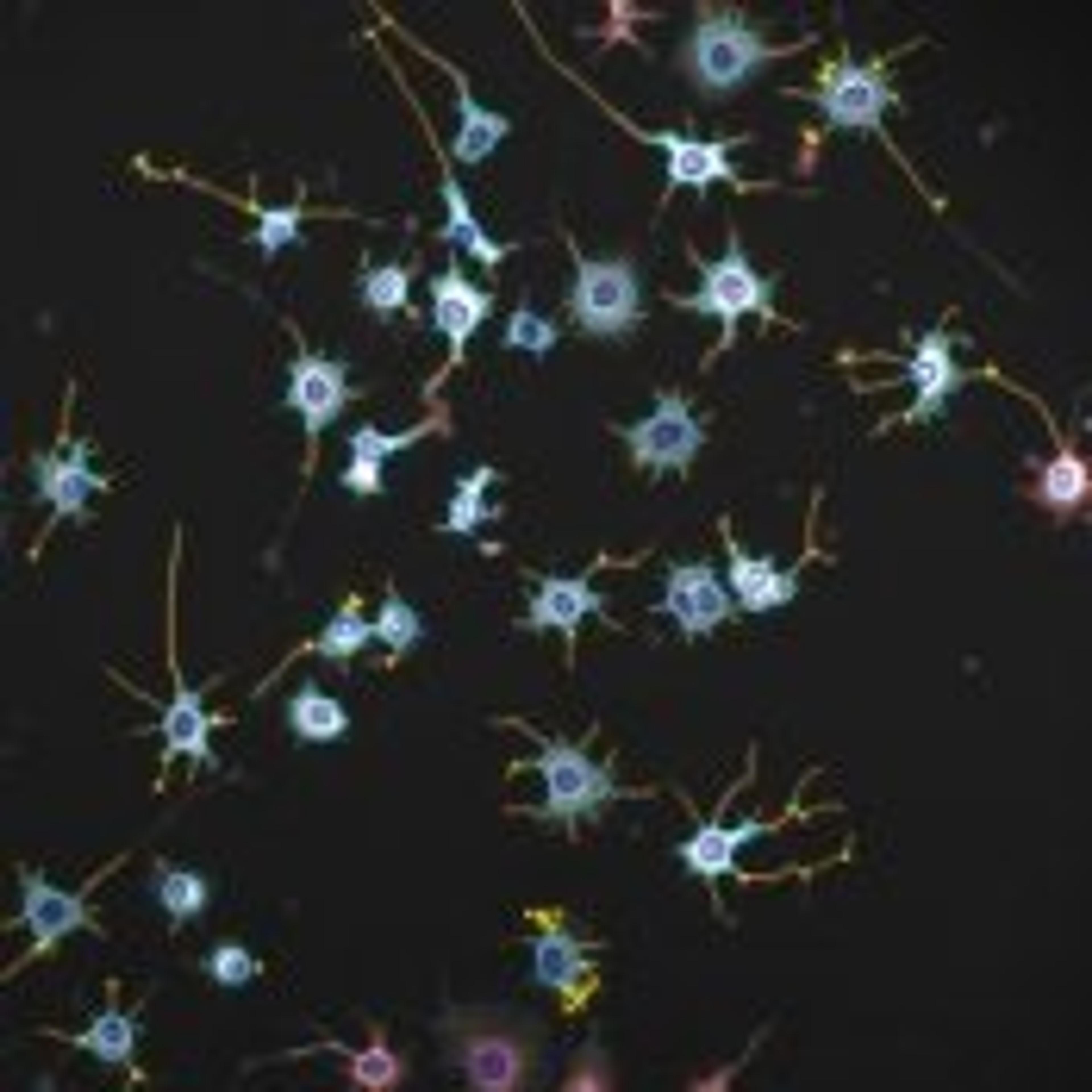
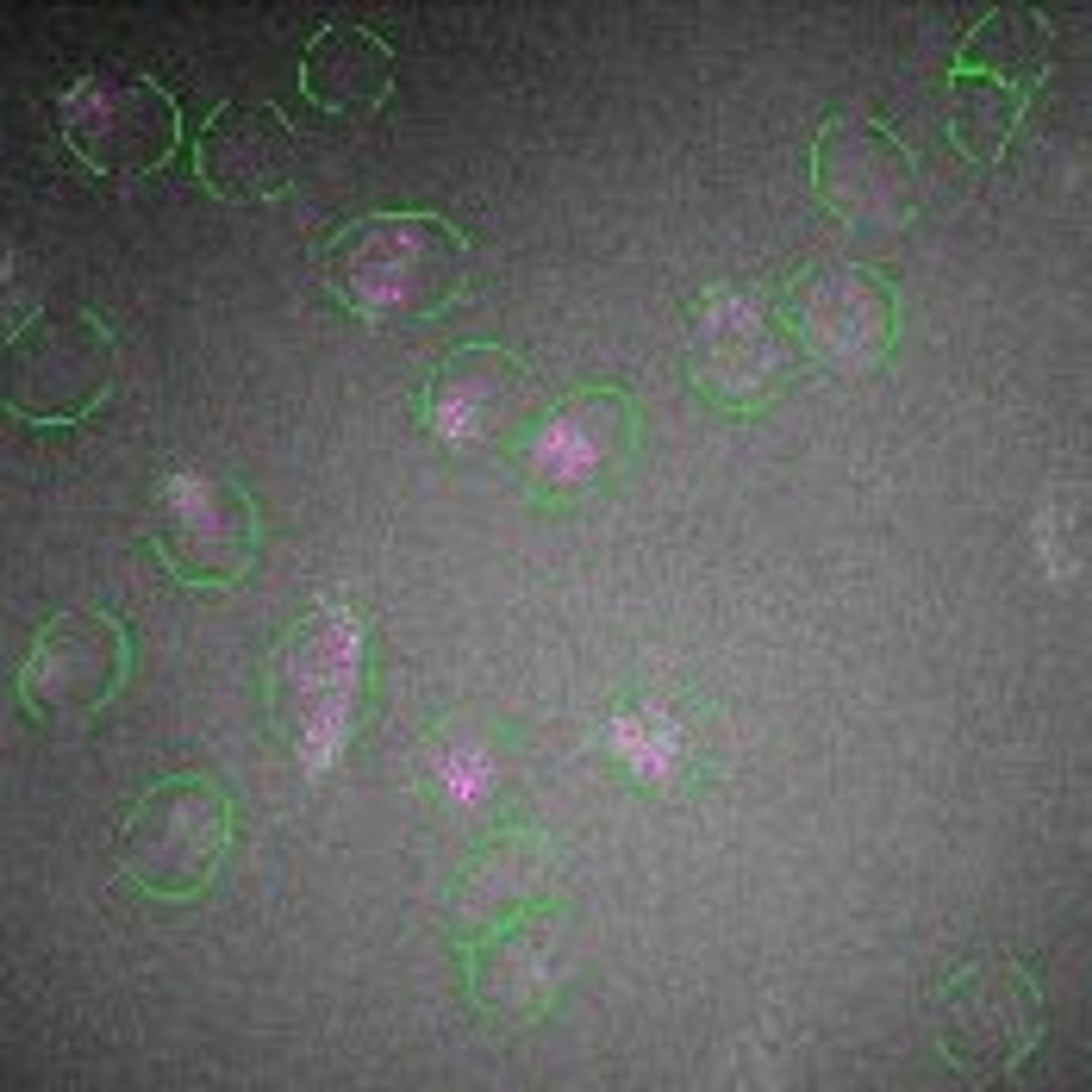
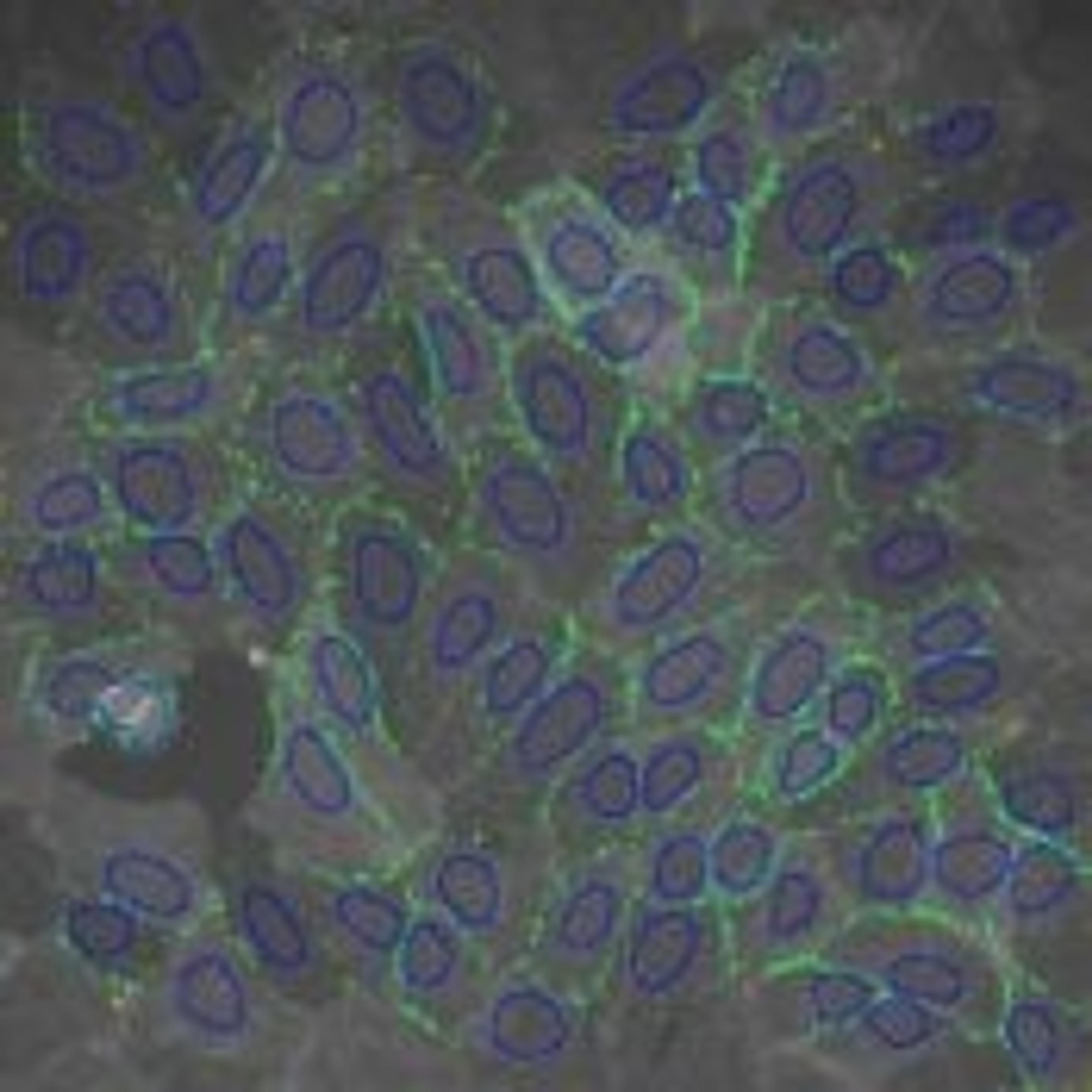
The Cell Cycle V2 BioApplication is a sophisticated image analysis algorithm that performs automated analysis of the cell cycle phase of individual and populations of cells. It reports cell cycle phase as well as the state of up to 3 target proteins for every cell analyzed, and correlates the target data with DNA content. This application has been biologically validated, enhancing confidence in your image-based data. Availa…
The Cytotoxicity V2 BioApplication is an image analysis application that simultaneously measures four markers of cytotoxicity (nuclear fragmentation/condensation, permeability, lysosomal mass-pH, and cell count) and reports individual and composite indicies of cytotoxicity. The V2 version has been enhanced with object segmentation to allow clumped cells to be efficiently analyzed. Available for the Cellomics ArrayScan VTI, A…
Extended Neurite Outgrowth V2 is an advanced image analysis algorithm that can quantitate neurite outgrowth with features such as cell body area and branch point quantitation, cell subpopulation analysis, Ability to analyze neuron subsets expressing up to two distinct neurotransmitters. It reports all features at the cell, well, and subpoulation level, and is biologically validated to enhance confidence in your image-based da…
Compartmental Analysis V2 is a powerful general image analysis tool for quantitating intracellular intensity changes in specific sub-cellular regions across multiple fluorescent channels. It performs spot analysis and reports intensity ratios and differences among sub-cellular regions and/or different fluorophores. This application has been biologically validated, enhancing confidence in your image-based data. Available for…
The Cell Motility V2 BioApplication is a advanced image analysis algorithm for measuring the overall motility of cells. It quantitates total distance traveled by each cell exposed to growth factor or compound for a quantiative assessment of cell motility. The V2 version includes advanced analytical capabilities such as population analysis and object segmentation. This application has been biologically validated, enhancing c…
Spot Detector V2 performs measurements of number, size, and other features of spots in cells. It is an easy-to-use and essential component of any image analysis software suite. This tool is excellent for measuring receptor internalization and trafficking, cell proliferation and counting, Analyzing populations. This application has been biologically validated, enhancing confidence in your image-based data. Available for the…
The Cell Spreading V2 BioApplication is an image analysis algorithm for measuring size and shape of cells. It utilizes cells labeled with cytotskeletal fluorescent reagents to report whether cells have rounded in response to compound treatment, and includes advanced features such as population characterization and object segmentation. This application has been biologically validated, enhancing confidence in your image-based…
The Neurite Outgrowth V2 BioApplication is a streamlined image analysis algorithm for morphological analysis of neurite outgrowth. Utilizes fluorescent images of neurons and neurites stained for tubulin to quantitate the number and length of neurites in a population of cells. This application has been biologically validated, enhancing confidence in your image-based data. Available for the Cellomics ArrayScan VTI, ArrayScan x…
Cyto-Cell Membrane Translocation V2 is a multi-channel image analysis algorithm for analysis of targets that translocate from cytoplasm to cell membrane. It utilizes colocalization of fluorescent markers to report whether the target is co-localized to the membrane, and reports cells' behavior relative to a control population. This application has been biologically validated, enhancing confidence in your image-based data. A…