RNAscope™ Multiplex Fluorescent Assays
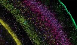
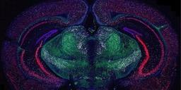
The RNAscope Multiplex Fluorescent assays are ideal for simultaneous detection studies of any genes in nearly any tissue-type using fluorescent labels.



The supplier does not provide quotations for this product through SelectScience. You can search for similar products in our Product Directory.
The RNAscope Multiplex Fluorescent assays are ideal for simultaneous detection studies of any genes in nearly any tissue-type using fluorescent labels. We offer with various levels of plexing to:
- FIND cells of interest by using cell specific markers
- CONFIRM cells by simultaneous detection of additional targets
- CHARACTERIZE cellular heterogeneity of complex tissues by mapping and identifying multiple cell subtypes simultaneously
Spatial analysis of gene expression is an essential tool for comprehensive studies of complex, highly heterogeneous tissues, such as the brain, tumors, and developing organs. Traditional RNA-ISH can be challenging due to low sensitivity, inconsistent detection accompanied with limited plexing. RNAscope Fluorescent Multiplex assays provide exceptional sensitivity, allowing single-molecule detection of RNA targets at the single cell level.
The multiplex assays offer exceptional sensitivity and allow single-molecule detection of up to 12 RNA targets simultaneously. These assays offer a novel and proprietary method of in situ hybridization (ISH) to achieve single cell resolution for RNA detection in a multitude of slide mounted samples. The assays are compatible with various sample types such as fresh frozen, fixed frozen and FFPE.
- Single molecule RNA detection.
- Single cell resolution
- Sensitive
- Specific
- Spatial Context

Spatial organization of multicellular immune hubs in human colorectal cancer
Immune responses to cancer are highly variable, with mismatch repair-deficient (MMRd) tumors exhibiting more anti-tumor immunity than mismatch repair-proficient (MMRp) tumors. To understand the rules governing these varied responses, researchers at the Broad Institute of MIT and Harvard transcriptionally profiled 371,223 cells from human colorectal tumors. Analysis revealed extensive transcriptional and spatial remodeling across tumors. To discover hubs of interacting malignant and immune cells, expression programs were identified in different cell types that co-varied across tumors from affected individuals and spatial profiling was used to localize coordinated programs. By identifying interacting cellular programs, the logic underlying spatially organized immune-malignant cell networks was revealed.
In this webinar, Dr. Jon Chen, Massachusetts General Hospital Cancer Center, Harvard Medical School, will discuss this study in greater detail and demonstrate a path to discovering multicellular interaction networks that underlie immunologic and tumorigenic processes in human cancer..
Key learning objectives
- How scRNA-seq can be used to reveal shared and distinct features of human MMRd and MMRp colorectal cancer
- How multicellular immune hubs can be predicted based on co-variation of gene programs
- Why understanding the molecular mechanisms underlying immune hubs and their temporal and spatial regulation upon treatment is critical for advancing cancer therapy
Certificate of attendance
All webinar participants can request a certificate of attendance, including a learning outcomes summary, for continuing education purposes.

Combining RNAscope™ Technology with Protein Detection: A Multi Omics Approach Using RNA-Protein Integrated Co-Detection Workflow (ICW)
The RNAscope in situ hybridization (ISH) technology is a powerful method for detecting gene expression with spatial and morphological tissue context. In this webinar, we will focus on our Integrated Co-Detection Workflow (ICW), which combines RNAscope ISH technology with immunohistochemistry (IHC) or immunofluorescence (IF) procedures to detect RNA and protein in the same sample. The webinar will cover guidelines for sample preparation, pretreatment, and detection, including tips for troubleshooting.
Key learning objectives
- Overview of RNAscope technologies
- Introduction to the Integrated Co-Detection Workflow (ICW)
- Guidelines on sample preparation and pretreatment
- Guidance on assay setup and appropriate controls
- Troubleshooting tips
- Examples of RNAscope co-detection results
Certificate of attendance
All webinar participants can request a certificate of attendance, including a learning outcomes summary, for continuing education purposes.
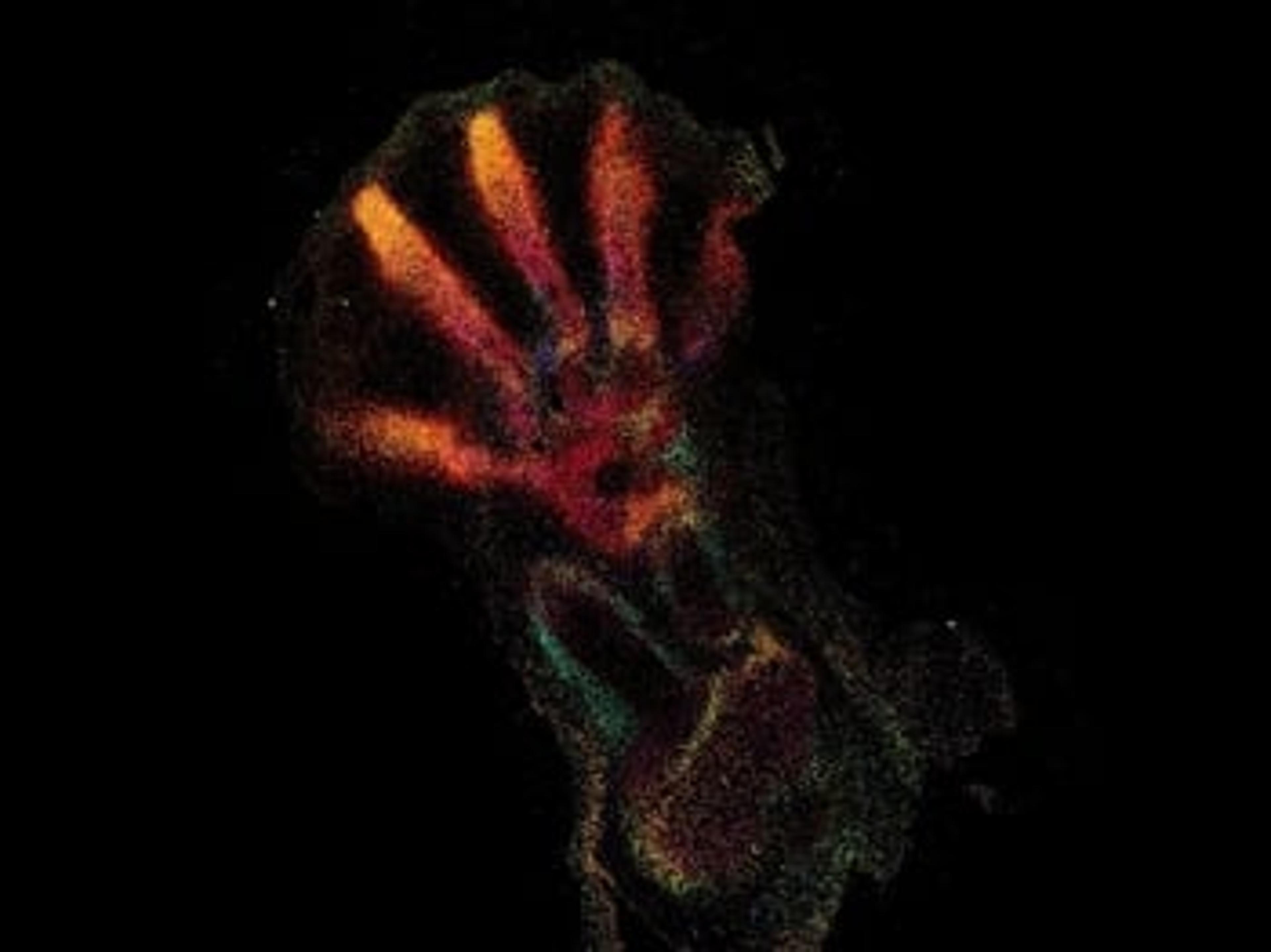
‘Google Maps’ for the human body – How spatial genomics is revolutionizing our understanding of disease
Discover how the Wellcome Sanger Institute is helping to develop a physical atlas of all human organs