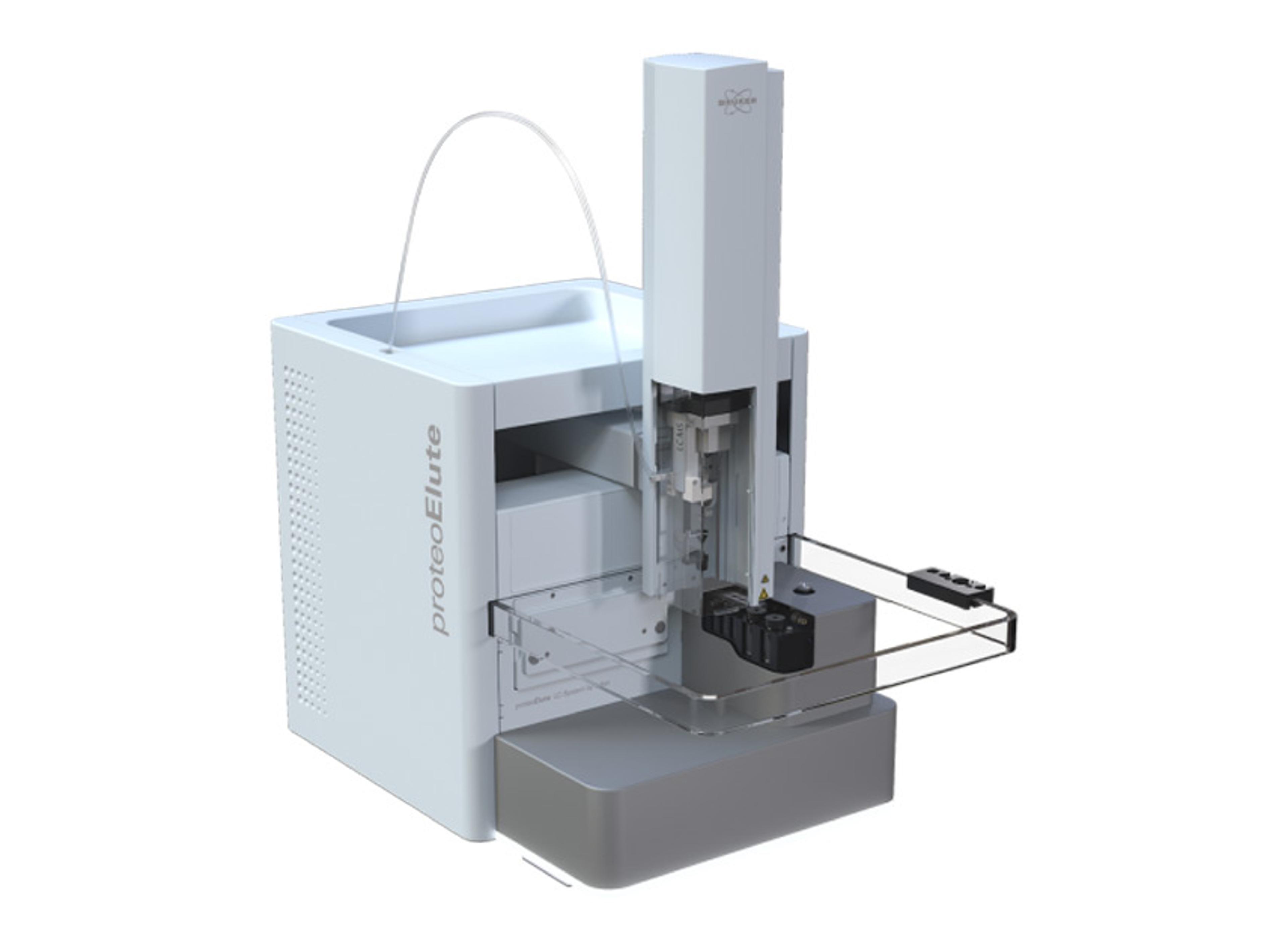
MMI CellCut
Clear-cut laser microdissection to isolate cells in tissue

MMI CellCut - Clear-cut Laser Microdissection

MMI CellCut - Clear-cut Laser Microdissection

Excising tissue with Laser Microdissection


The supplier does not provide quotations for this product through SelectScience. You can search for similar products in our Product Directory.
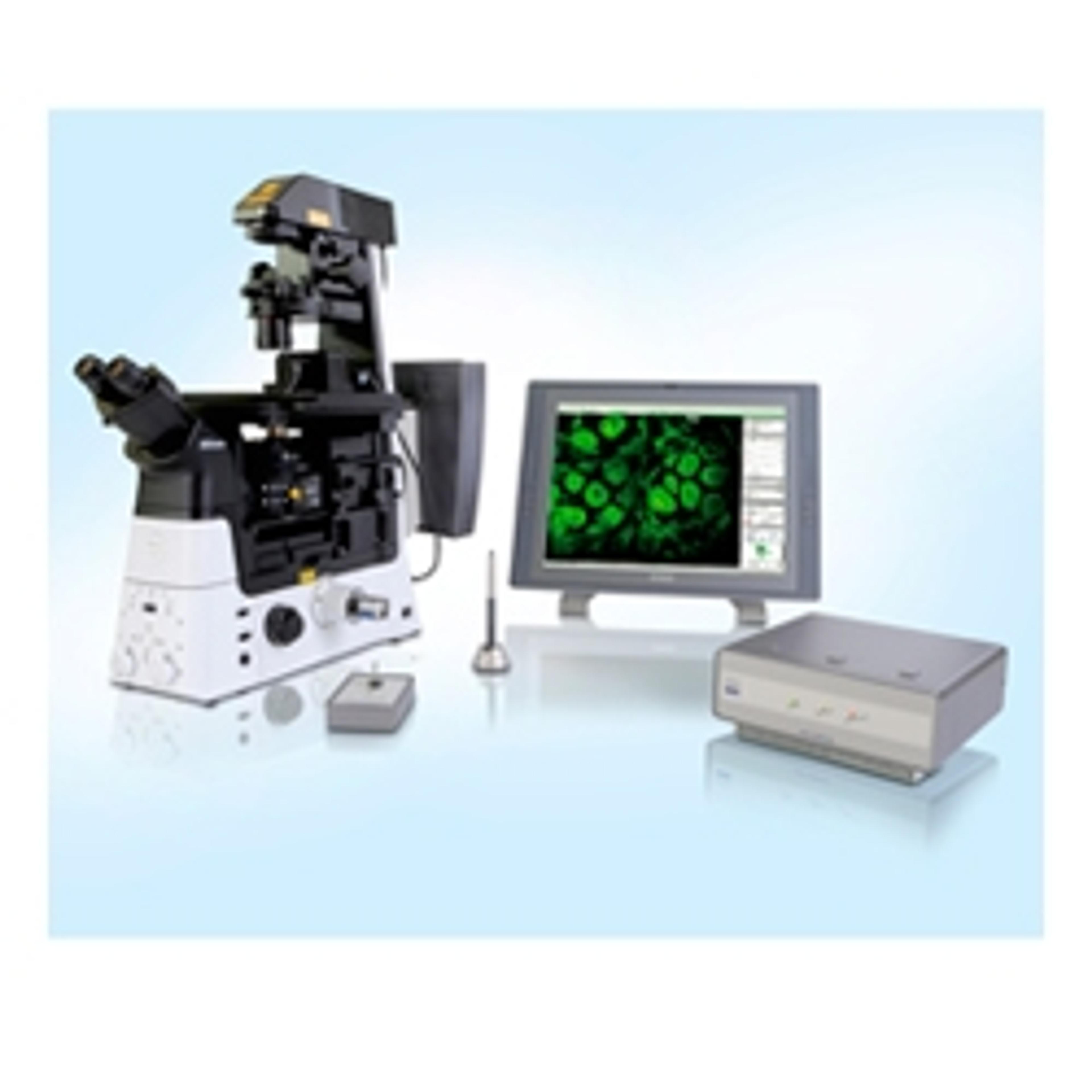
The MMI CellCut facilitates precise and contamination-free dissection of cell clusters, single cells or sub-cellular compartments from various types of tissues including fresh frozen, paraffin embedded and archived slides, cytospins, smears and even living cells.
Laser Microdissection is a well-established technology to cut cells from tissue when the separate analysis of specific subsets of your sample is crucial to obtain relevant and significant data.
The MMI CellCut uses a precisely focused UV-Laser for clear-cut results. To obtain a homogeneous cell population for meaningful downstream analysis, the unique MMI CapLift technology provides gentle, contamination-free and highly efficient sample collection method including full visual inspection at any step during and after isolation. The MMI CellCut workflow is fully compatible with most downstream analyses enabling highly reliable analysis and diagnosis.
Features and Benefits:
- Precise cutting of various tissue types and even living cells using patented technology
- Flexible and quick sample selection, inspection and precise positioning of cut cells by unique and fully auditable Predefined Target Positioning (PTP) feature
- Contamination-free and gentle sample capture with 8 different types of MMI collection tubes and MMI MultiCap strips
- Fully compatible with downstream analysis employing unstained samples and low damage laser
Applications
The MMI CellCut can be applied in various different research projects:
- when the separate analysis of specific subsets of your sample is crucial to obtain relevant and significant data
- when the precise and contamination-free isolation of cells from tissue is required
Typical applications include:
- Cancer Research
- Oncology
- Molecular Pathology
- Cell Biology
- Botany and Plant Research
- Forensic Analysis

Stress disrupts reward in mice: insights into neurocircuits and molecular targets in psychiatry
Reduced sensitivity to reward is common in stress-related neuropsychiatric disorders, including symptoms such as anhedonia and apathy in depression and schizophrenia, for example. Animal models are essential to increase understanding of the underlying neurobiology.
In this webinar, Professor Christopher Pryce, Professor of Neurobiology and Biological Psychiatry at the University of Zurich, will explore the effects of chronic stress on reward behavior and associated activity in the glutamate neuron pathway from basal amygdala to nucleus accumbens (BA-NAc). Furthermore, he will discuss the importance of molecular tools, in particular laser capture microdissection for the identification and collection of BA-NAc neuron populations for RNA-sequencing.
Additionally, he will explain how further research, focused on specific subpopulations of BA-NAc neurons, may lead to identifying potential targets for novel treatments of reward-related pathologies in neuropsychiatric disorders.
Key learning objectives
- Learn about stress effects on reward behavior and BA-NAc neural pathway activity in mice
- Explore the process of replicating stress effects on reward by manipulating the activity of BA-NAc neurons
- Understand the importance of laser capture microdissection to study specific neuronal populations
- Learn about RNA-sequencing with LCM-collected neurons
Who should attend?
- Researchers interested in vivo-ex vivo questions in behavioural neurobiology
- Researchers interested in studying -omics in specific (brain) cell populations
Certificate of attendance
All webinar participants can request a certificate of attendance, including a learning outcomes summary, for continuing education purposes.
MMI CellCut
In this video, MMI Molecular Machines & Industries presents the MMI CellCut which utilizes clear-cut laser microdissection to isolate cells in tissue. The MMI CellCut facilitates precise and contamination-free dissection of cell clusters, single cells, or sub-cellular compartments from various types of tissues including fresh frozen, paraffin embedded and archived slides, cytospins, smears, and even living cells.