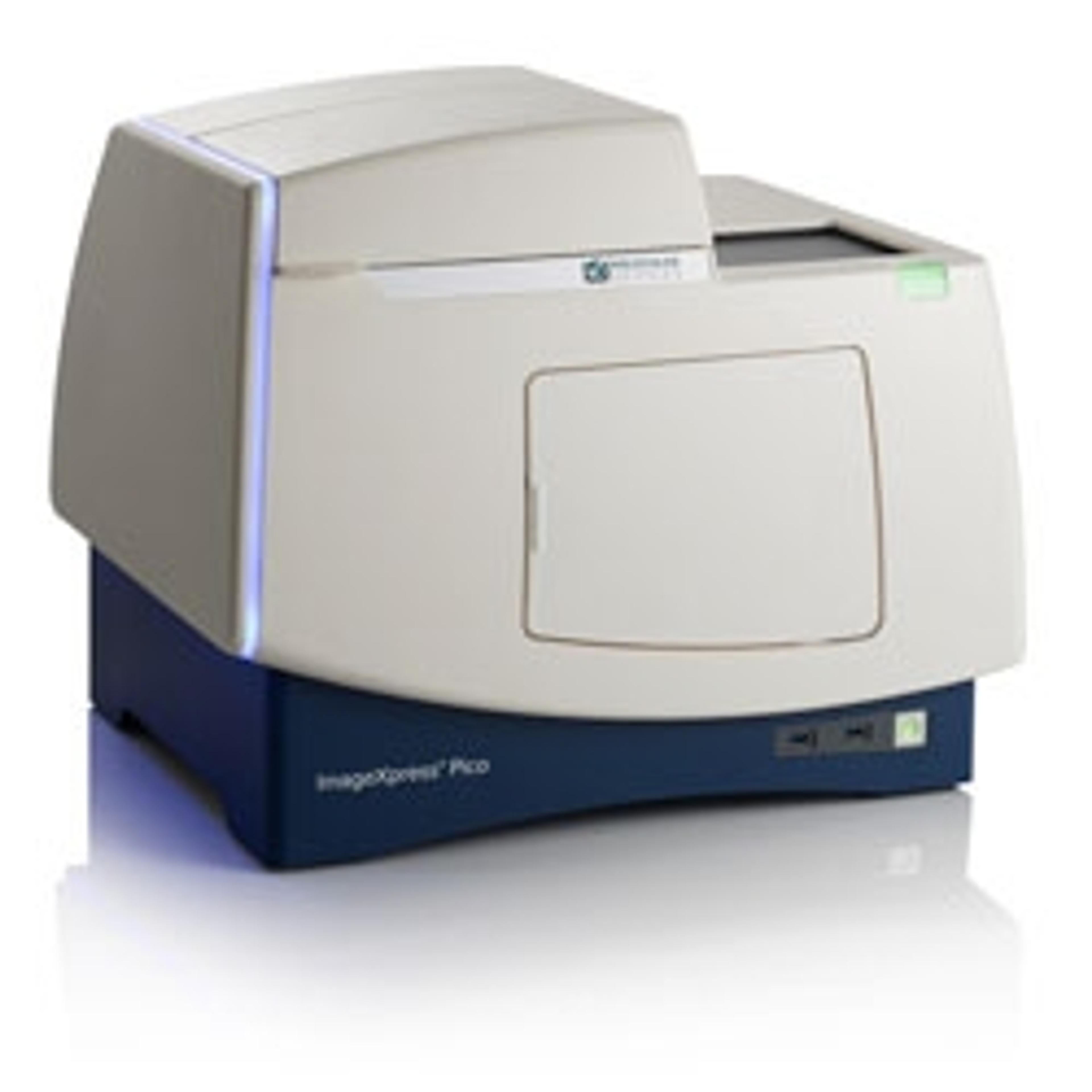
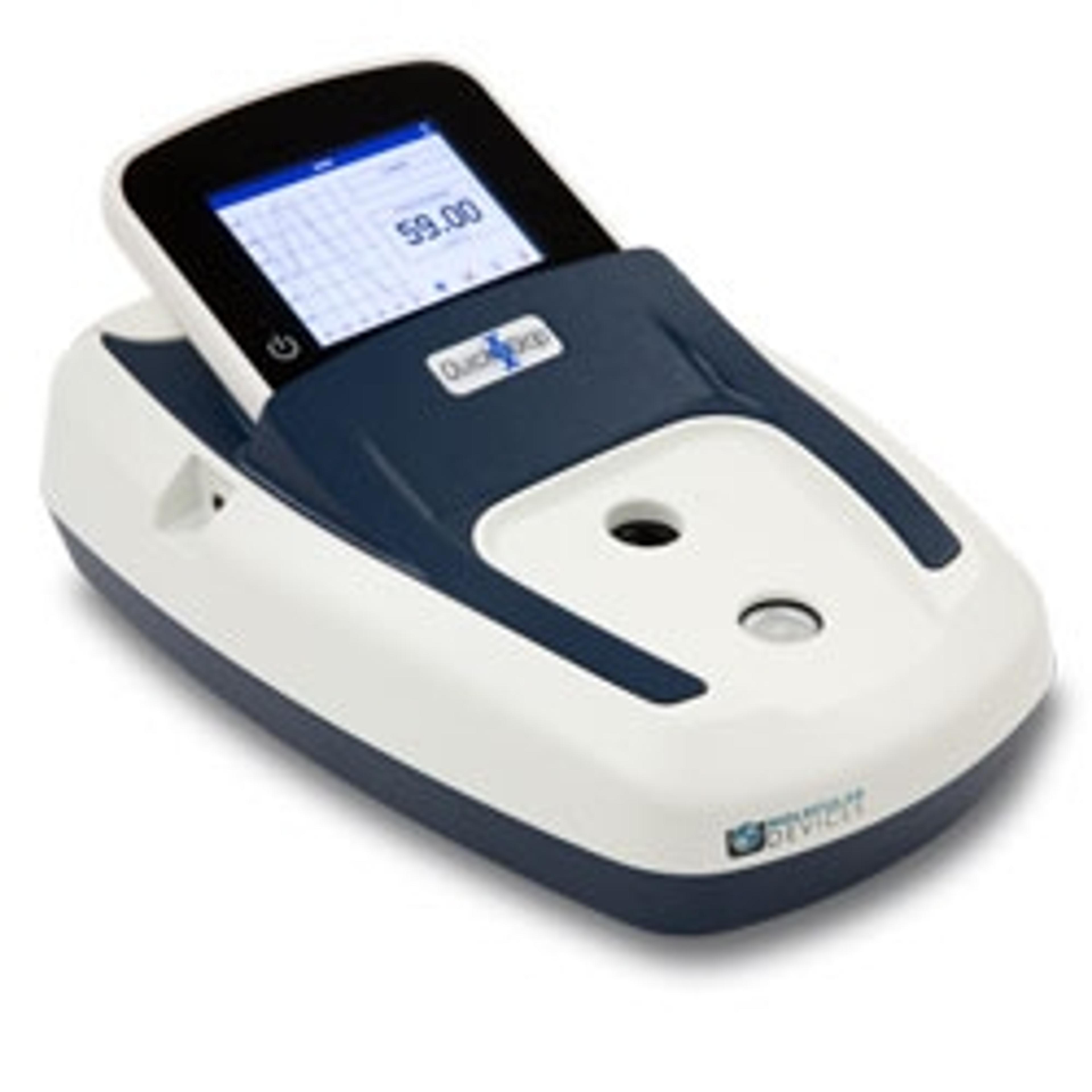
ImageXpress Pico Automated Cell Imaging System
Affordable cellular imaging and analysis system for individual biology labs - go from sample to results in minutes



Receive your quote directly from the manufacturer.
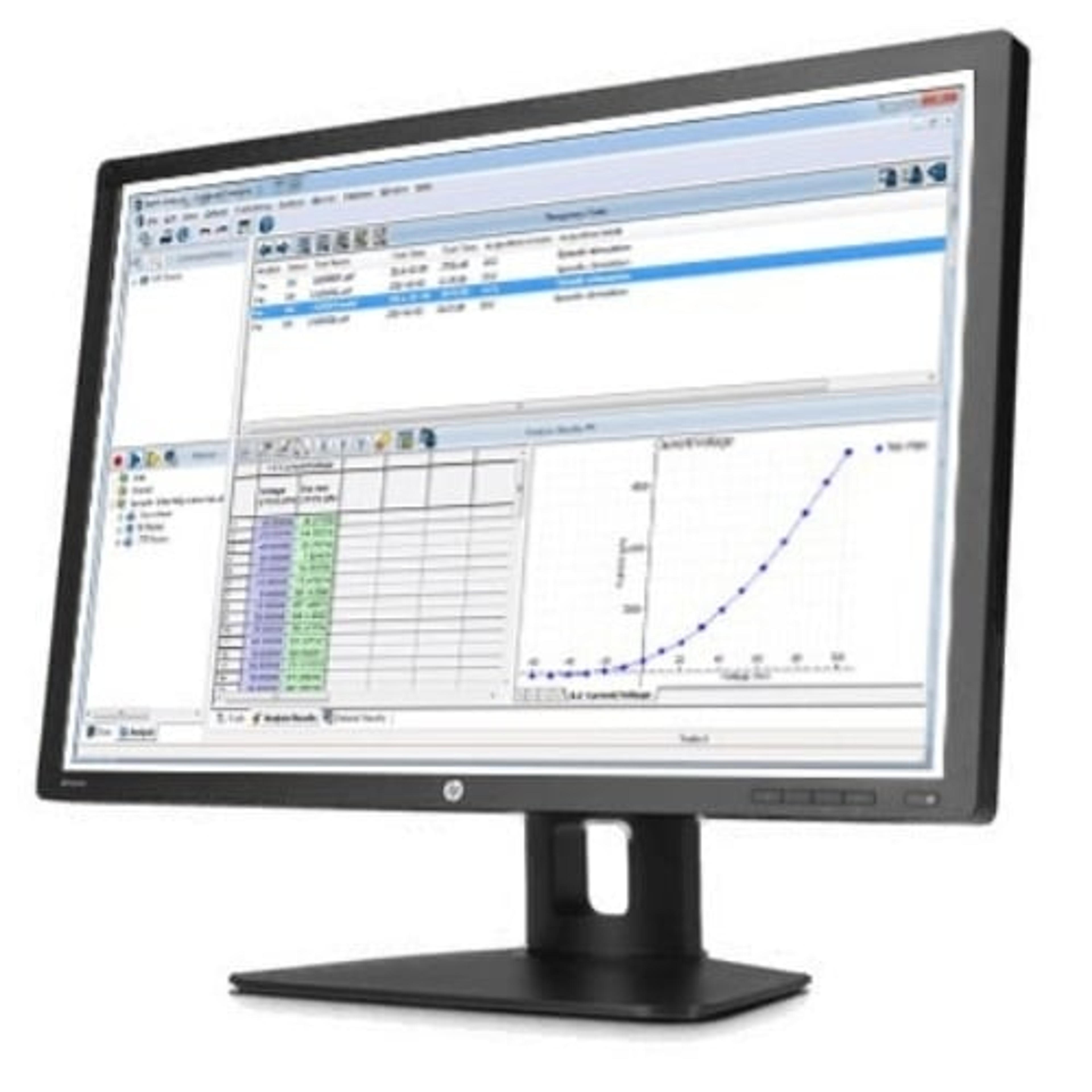
The ImageXpress® Pico Automated Cell Imaging System combines high-resolution imaging and powerful analysis, with a streamlined workflow. It includes CellReporterXpress® software for acquisition and analysis, and features a comprehensive portfolio of preconfigured protocols. The automated setup of analysis parameters shortens the learning curve, so you can start running experiments quickly. If you’re in the market for fluorescence imaging or digital microscopy, the ease and affordability of an automated imager is now within reach.

Cellular imaging insights
The goal of many researchers is to elucidate the complex cellular pathways within 2D and 3D cellular structures in response to chemical stimulation or stress. The capacity to which scientists have access to and can adopt emerging cellular imaging and analysis technology fuels their level of understanding. In this eBook, Molecular Devices provides an overview of the new tools that can be used to better understand complex imaging models.

Human organoid lines for modelling the intestinal epithelial barrier in vitro
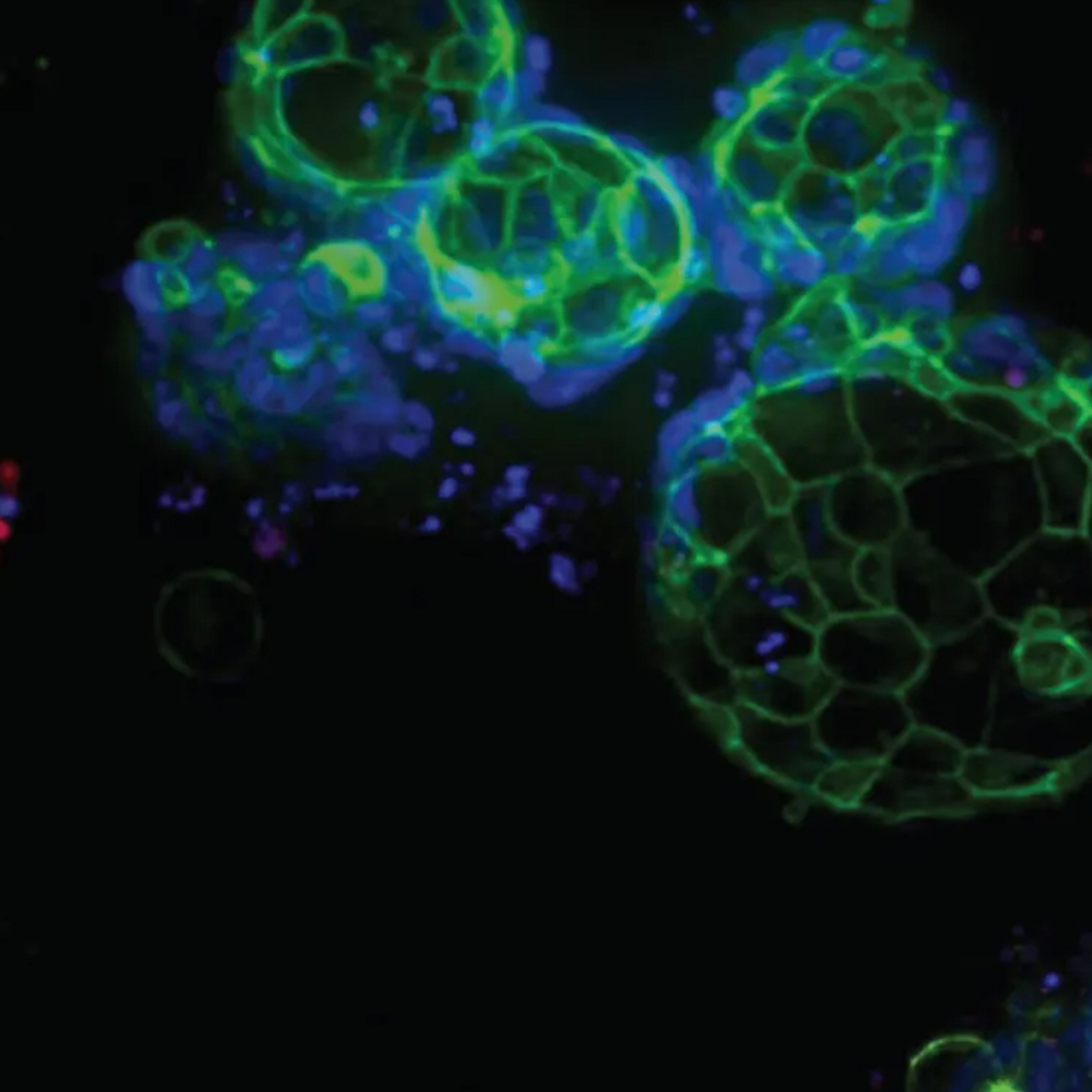
In this application note, epithelial monolayers were derived from two 3D-Ready non-cancerous human duodenal organoid lines from Molecular Devices and morphological and functional analyses were conducted to assess the resultant monolayers' ability to model the human intestinal barrier.

Transform your cell imaging
From endpoint and live-cell imaging applications to imaging and analyzing 2D monolayers, 3D cell models, and organ-on-a-chip platforms, the world of cellular imaging is very diverse. This eBook highlights the use of the ImageXpress® Pico Automated Cell Imaging System and CellReporterXpress® Automated Image Acquisition and Analysis Software to perform a wide variety of complex cellular imaging assays easily.