ZEISS Video Interviews: Hear from the Material Science Experts
Watch these exclusive video interviews regarding the use of state-of-the-art microscopy for materials analysis
4 Nov 2016
Editorial article
Dr Uros Krzic, application consultant at Carl ZEISS Microscopy discusses high resolution confocal microscopy
In these video interviews, learn about the use of confocal microscopy in material sciences, discover more about how ZEISS microscopy is being used in nanomaterial research, and find out about the development of complex 3D nano-structures.
1. Optimizing Energy Conversion and Storage Materials Using Correlative Microscopy at HZB
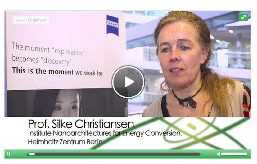
Professor Christiansen discusses nanomaterials for solar cells and energy storage
Professor Silke Christiansen, Professor at Frei University in Berlin, discusses her research at Helmholtz Center for Materials and Energy into nanomaterials for solar cells and energy storage. She is studying new ways of converting solar energy into other forms of energy as well as storing energy in an efficient way.
2. Confocal Microscopy in Materials Science
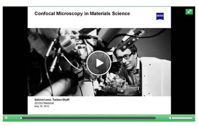
Learn about the use of confocal microscopy in this webinar
In this webinar, Sabine Lenz discusses using confocal microscopy in materials science with Torben Wulff from ZEISS Microscopy, giving examples of applications and the different microscopes used.
3. Presentation: Correlative Microscopy-Spectroscopy Studies of Complex 3D Nano-Structures
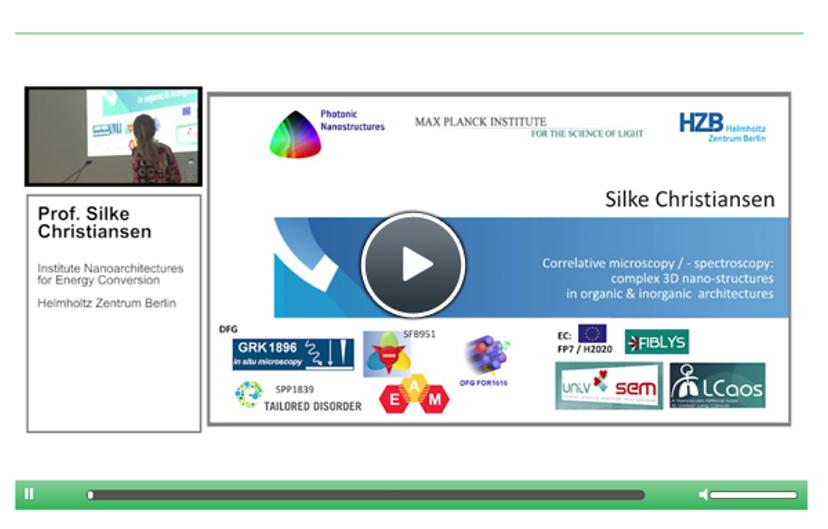
Find out more about 3D nano-structures in this presentation
In this presentation, Professor Silke Christiansen, Helmholtz-Zentrum Berlin für Materialien und Energie (HZB), discusses her research into the development of energy efficient materials with complex 3D nano-structures. Hear how her lab is collaborating with ZEISS microscopy to create innovative solutions combining correlative microscopy and spectroscopy for the detailed analysis organic and inorganic materials. Professor Christiansen demonstrates the benefits of this technique for the analysis of human bone and other medical samples.
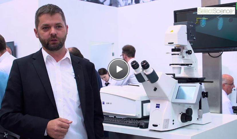
4. Enhanced Speed for Sensitive, High Resolution Confocal Microscopy

Dr Krzic discusses transient event imaging in this interview
Dr Uros Krzic, application consultant at Carl ZEISS Microscopy GmbH, describes how the ZEISS Airyscan with fast acquisition mode facilitates sensitive, high resolution confocal microscopy that is ideal for imaging transient events.
Related products
Request Quote for All Products
ZEISS MultiSEM Research Partner Program
ZEISS Research Microscopy SolutionsThe World's Fastest Scanning Electron Microscopes.
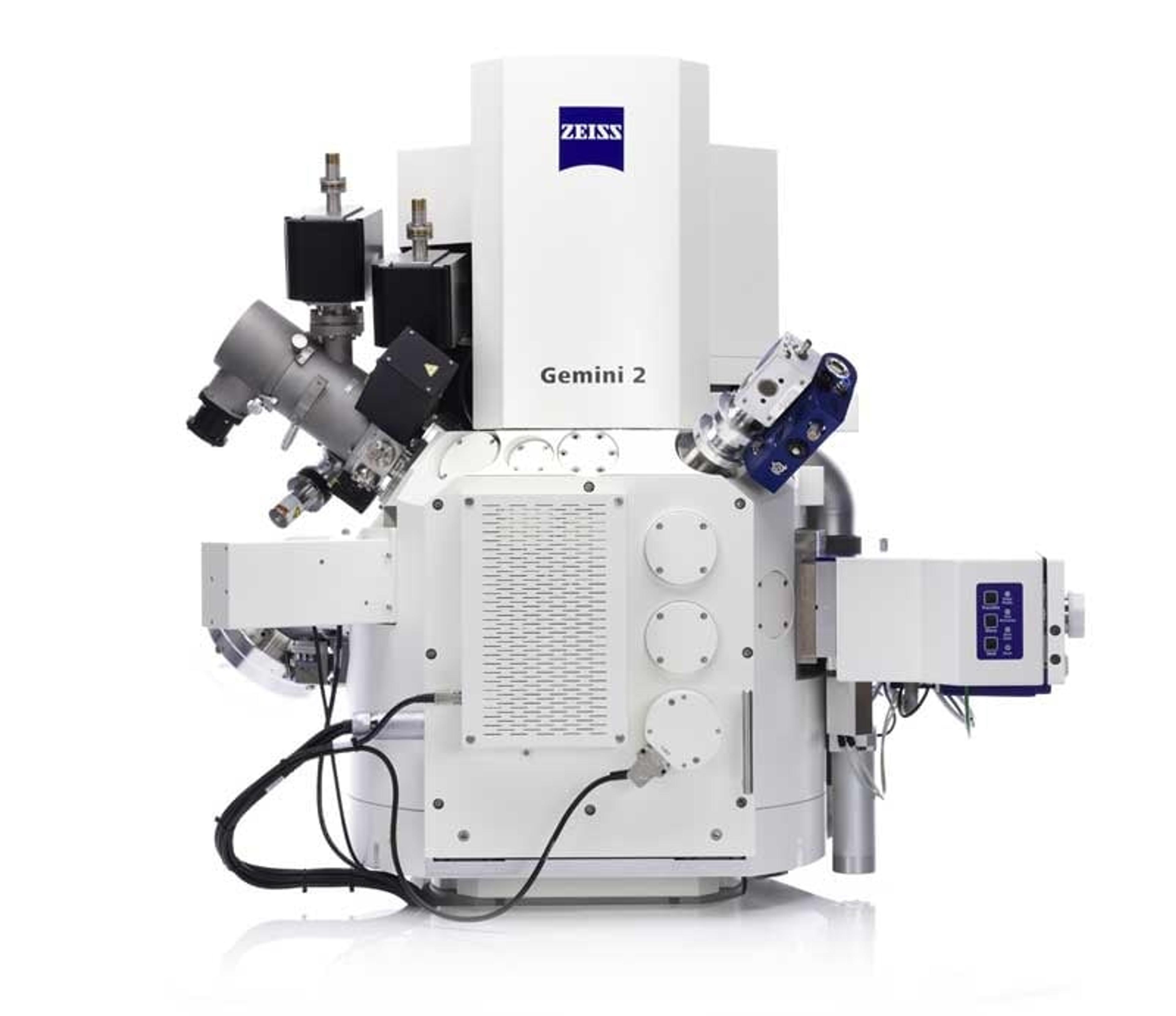
ZEISS Crossbeam Family
ZEISS Research Microscopy SolutionsWithin ZEISS Crossbeam Family you have the choice between Crossbeam 340 or Crossbeam 550. Exploit the variable pressure capabilities of Crossbeam 340. Or use Crossbeam 550 for your most demanding characterizations and choose the chamber size, standard or large, that best suits your samples.